 Movie
Movie Controller

[English] 日本語
 Yorodumi
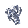
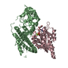
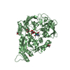
- PDB-4ieb: Crystal Structure of a Gly128Met mutant of the toxoplasma CDPK, T... -
+ Open data

- Basic information
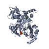
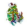
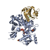
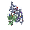
| Entry | Database: PDB / ID: 4ieb | ||||||
|---|---|---|---|---|---|---|---|
| Title | Crystal Structure of a Gly128Met mutant of the toxoplasma CDPK, TGME49_101440 | ||||||
 Components Components | Calmodulin-domain protein kinase 1 | ||||||
 Keywords Keywords |  TRANSFERASE / cdpks / toxoplasma / kinase / protist / Structural Genomics / Structural Genomics Consortium / SGC / ATP-binding / Nucleotide-binding / Serine/threonine-protein kinase TRANSFERASE / cdpks / toxoplasma / kinase / protist / Structural Genomics / Structural Genomics Consortium / SGC / ATP-binding / Nucleotide-binding / Serine/threonine-protein kinase | ||||||
| Function / homology |  Function and homology informationphosphorylation / protein serine/threonine kinase activity / calcium ion binding / ATP binding / membrane / cytoplasm Function and homology informationphosphorylation / protein serine/threonine kinase activity / calcium ion binding / ATP binding / membrane / cytoplasmSimilarity search - Function | ||||||
| Biological species |  Toxoplasma gondii (eukaryote) Toxoplasma gondii (eukaryote) | ||||||
| Method | X-RAY DIFFRACTION / SYNCHROTRON / MOLECULAR REPLACEMENT / molecular replacement / Resolution: 2.05 Å | ||||||
 Authors Authors | Wernimont, A.K. / Artz, J.D. / El Bakkouri, M. / Schapira, M. / Edwards, A.M. / Arrowsmith, C.H. / Bountra, C. / Hui, R. / Amani, M. / Structural Genomics Consortium (SGC) | ||||||
 Citation Citation | Journal: To be Published Title: Crystal Structure of a Gly128Met mutant of the toxoplasma CDPK, TGME49_101440 Authors: Wernimont, A.K. / Artz, J.D. / El Bakkouri, M. / Schapira, M. / Edwards, A.M. / Arrowsmith, C.H. / Bountra, C. / Hui, R. / Amani, M. | ||||||
| History |
|
- Structure visualization
| Structure viewer | Molecule: MolmilJmol/JSmol |
|---|
- Downloads & links
-Download
| PDBx/mmCIF format | 4ieb.cif.gz | 204.1 KB | Display | PDBx/mmCIF format |
|---|---|---|---|---|
| PDB format | pdb4ieb.ent.gz | 168.2 KB | Display | PDB format |
| PDBx/mmJSON format | 4ieb.json.gz | Tree view | PDBx/mmJSON format | |
| Others |  Other downloads Other downloads |
-Validation report
| Arichive directory | https://data.pdbj.org/pub/pdb/validation_reports/ie/4iebftp://data.pdbj.org/pub/pdb/validation_reports/ie/4ieb | HTTPS FTP |
|---|
-Related structure data
| Similar structure data |
|---|
-Links
 PDBj
PDBj


- Assembly
| Deposited unit | 
| ||||||||
|---|---|---|---|---|---|---|---|---|---|
| 1 |
| ||||||||
| Unit cell |
|
-Components
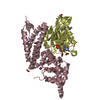
| #1: Protein | Mass: 58478.352 Da / Num. of mol.: 1 / Mutation: G128M Source method: isolated from a genetically manipulated source Source: (gene. exp.) Toxoplasma gondii (eukaryote) / Gene: CDPK1, TGGT1_059880, TGME49_101440, TGVEG_042030 / Production host:  Escherichia coli (E. coli) Escherichia coli (E. coli)References: UniProt: Q9BJF5, Ca2+/calmodulin-dependent protein kinase | ||||
|---|---|---|---|---|---|
| #2: Chemical | ChemComp-CA /   Mass: 40.078 Da / Num. of mol.: 4 / Source method: obtained synthetically / Formula: Ca Mass: 40.078 Da / Num. of mol.: 4 / Source method: obtained synthetically / Formula: Ca#3: Chemical | ChemComp-ANP / |   Mass: 506.196 Da / Num. of mol.: 1 / Source method: obtained synthetically / Formula: C10H17N6O12P3 / Comment: AMP-PNP, energy-carrying molecule analogue*YM Mass: 506.196 Da / Num. of mol.: 1 / Source method: obtained synthetically / Formula: C10H17N6O12P3 / Comment: AMP-PNP, energy-carrying molecule analogue*YM#4: Water | ChemComp-HOH / | Water Mass: 18.015 Da / Num. of mol.: 321 / Source method: isolated from a natural source / Formula: H2O Mass: 18.015 Da / Num. of mol.: 321 / Source method: isolated from a natural source / Formula: H2O |
-Experimental details
-Experiment
| Experiment | Method: X-RAY DIFFRACTION / Number of used crystals: 1 |
|---|
- Sample preparation
| Crystal | Density Matthews: 2.12 Å3/Da / Density % sol: 42.06 % |
|---|---|
| Crystal grow | Temperature: 293 K / Method: vapor diffusion, sitting drop / pH: 9.5 Details: 14% PEG3350, 0.3 M MgAcetate, 0.1 M Glycine pH 9.5, 5 mM CaCl2, 2 mM MgCl2, 5 mM AMPPNP, VAPOR DIFFUSION, SITTING DROP, temperature 293K |
-Data collection
| Diffraction | Mean temperature: 100 K | |||||||||||||||||||||||||||||||||||||||||||||||||||||||||||||||||||||||||||||||||||||||||||||||||||||||||||||||||||||||||||||||||||||||||||||||||||
|---|---|---|---|---|---|---|---|---|---|---|---|---|---|---|---|---|---|---|---|---|---|---|---|---|---|---|---|---|---|---|---|---|---|---|---|---|---|---|---|---|---|---|---|---|---|---|---|---|---|---|---|---|---|---|---|---|---|---|---|---|---|---|---|---|---|---|---|---|---|---|---|---|---|---|---|---|---|---|---|---|---|---|---|---|---|---|---|---|---|---|---|---|---|---|---|---|---|---|---|---|---|---|---|---|---|---|---|---|---|---|---|---|---|---|---|---|---|---|---|---|---|---|---|---|---|---|---|---|---|---|---|---|---|---|---|---|---|---|---|---|---|---|---|---|---|---|---|---|
| Diffraction source | Source: SYNCHROTRON / Site: APS  / Beamline: 19-ID / Wavelength: 0.97935 Å / Beamline: 19-ID / Wavelength: 0.97935 Å | |||||||||||||||||||||||||||||||||||||||||||||||||||||||||||||||||||||||||||||||||||||||||||||||||||||||||||||||||||||||||||||||||||||||||||||||||||
| Detector | Type: ADSC QUANTUM 315 / Detector: CCD / Date: Feb 17, 2010 | |||||||||||||||||||||||||||||||||||||||||||||||||||||||||||||||||||||||||||||||||||||||||||||||||||||||||||||||||||||||||||||||||||||||||||||||||||
| Radiation | Protocol: SINGLE WAVELENGTH / Monochromatic (M) / Laue (L): M / Scattering type: x-ray | |||||||||||||||||||||||||||||||||||||||||||||||||||||||||||||||||||||||||||||||||||||||||||||||||||||||||||||||||||||||||||||||||||||||||||||||||||
| Radiation wavelength | Wavelength: 0.97935 Å / Relative weight: 1 | |||||||||||||||||||||||||||||||||||||||||||||||||||||||||||||||||||||||||||||||||||||||||||||||||||||||||||||||||||||||||||||||||||||||||||||||||||
| Reflection | Resolution: 2.05→50 Å / Num. all: 32213 / Num. obs: 31956 / % possible obs: 99.2 % / Observed criterion σ(F): 0 / Observed criterion σ(I): 0 / Redundancy: 7.1 % / Biso Wilson estimate: 22.5 Å2 / Rmerge(I) obs: 0.06 / Χ2: 1.383 / Net I/σ(I): 12.5 | |||||||||||||||||||||||||||||||||||||||||||||||||||||||||||||||||||||||||||||||||||||||||||||||||||||||||||||||||||||||||||||||||||||||||||||||||||
| Reflection shell | Diffraction-ID: 1
|
-Phasing
| Phasing | Method: molecular replacement | |||||||||
|---|---|---|---|---|---|---|---|---|---|---|
| Phasing MR | Rfactor: 36.57 / Model details: Phaser MODE: MR_AUTO
|
- Processing
| Software |
| |||||||||||||||||||||||||||||||||||||||||||||
|---|---|---|---|---|---|---|---|---|---|---|---|---|---|---|---|---|---|---|---|---|---|---|---|---|---|---|---|---|---|---|---|---|---|---|---|---|---|---|---|---|---|---|---|---|---|---|
| Refinement | Method to determine structure: MOLECULAR REPLACEMENT / Resolution: 2.05→48.39 Å / Cor.coef. Fo:Fc: 0.955 / Cor.coef. Fo:Fc free: 0.934 / WRfactor Rfree: 0.2151 / WRfactor Rwork: 0.1722 / Occupancy max: 1 / Occupancy min: 0.5 / FOM work R set: 0.8523 / SU B: 8.072 / SU ML: 0.114 / SU R Cruickshank DPI: 0.2011 / SU Rfree: 0.1679 / Cross valid method: THROUGHOUT / σ(F): 0 / ESU R: 0.201 / ESU R Free: 0.168 / Stereochemistry target values: MAXIMUM LIKELIHOOD / Details: HYDROGENS HAVE BEEN USED IF PRESENT IN THE INPUT
| |||||||||||||||||||||||||||||||||||||||||||||
| Solvent computation | Ion probe radii: 0.8 Å / Shrinkage radii: 0.8 Å / VDW probe radii: 1.2 Å / Solvent model: MASK | |||||||||||||||||||||||||||||||||||||||||||||
| Displacement parameters | Biso max: 73.9 Å2 / Biso mean: 27.759 Å2 / Biso min: 6.69 Å2
| |||||||||||||||||||||||||||||||||||||||||||||
| Refinement step | Cycle: LAST / Resolution: 2.05→48.39 Å
| |||||||||||||||||||||||||||||||||||||||||||||
| Refine LS restraints |
| |||||||||||||||||||||||||||||||||||||||||||||
| LS refinement shell | Resolution: 2.05→2.103 Å / Total num. of bins used: 20
| |||||||||||||||||||||||||||||||||||||||||||||
| Refinement TLS params. | Method: refined / Origin x: 20.995 Å / Origin y: -0.9 Å / Origin z: -6.9889 Å
| |||||||||||||||||||||||||||||||||||||||||||||
| Refinement TLS group |
|
