 Movie
Movie Controller

[English] 日本語
 Yorodumi
- PDB-3rw0: Crystal structure of the NavAb voltage-gated sodium channel (Met2... -
+ Open data

- Basic information
| Entry | Database: PDB / ID: 3rw0 | ||||||
|---|---|---|---|---|---|---|---|
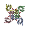
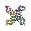
| Title | Crystal structure of the NavAb voltage-gated sodium channel (Met221Cys, 2.95 A) | ||||||
 Components Components | Ion transport protein Ion transporter Ion transporter | ||||||
 Keywords Keywords | METAL TRANSPORT / tetrameric ion channel / voltage-gated sodium-selective ion channel / membrane | ||||||
| Function / homology |  Function and homology information Function and homology informationmonoatomic cation channel activity / identical protein binding / plasma membraneSimilarity search - Function | ||||||
| Biological species |  Arcobacter butzleri (bacteria) Arcobacter butzleri (bacteria) | ||||||
| Method | X-RAY DIFFRACTION / SYNCHROTRON / MOLECULAR REPLACEMENT / Resolution: 2.95 Å | ||||||
 Authors Authors | Payandeh, J. / Scheuer, T. / Zheng, N. / Catterall, W.A. | ||||||
 Citation Citation | Journal: Nature / Year: 2011 Title: The crystal structure of a voltage-gated sodium channel. Authors: Payandeh, J. / Scheuer, T. / Zheng, N. / Catterall, W.A. | ||||||
| History |
|
- Structure visualization
| Structure viewer | Molecule: MolmilJmol/JSmol |
|---|
- Downloads & links
-Download
| PDBx/mmCIF format | 3rw0.cif.gz | 106.6 KB | Display | PDBx/mmCIF format |
|---|---|---|---|---|
| PDB format | pdb3rw0.ent.gz | 85.3 KB | Display | PDB format |
| PDBx/mmJSON format | 3rw0.json.gz | Tree view | PDBx/mmJSON format | |
| Others |  Other downloads Other downloads |
-Validation report
| Arichive directory | https://data.pdbj.org/pub/pdb/validation_reports/rw/3rw0ftp://data.pdbj.org/pub/pdb/validation_reports/rw/3rw0 | HTTPS FTP |
|---|
-Related structure data


-Links
 PDBj
PDBj

- Assembly
| Deposited unit | 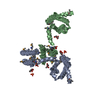
| ||||||||
|---|---|---|---|---|---|---|---|---|---|
| 1 | 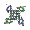
| ||||||||
| Unit cell |
|
-Components
| #1: Protein | Ion transporter Mass: 33649.789 Da / Num. of mol.: 2 / Mutation: M221C Source method: isolated from a genetically manipulated source Source: (gene. exp.) Arcobacter butzleri (bacteria) / Strain: RM4018 / Gene: Abu_1752 / Plasmid: pFastBac Dual / Production host:  Trichoplusia ni (cabbage looper) / Strain (production host): Hi5 / References: UniProt: A8EVM5 Trichoplusia ni (cabbage looper) / Strain (production host): Hi5 / References: UniProt: A8EVM5#2: Chemical | ChemComp-PX4 / Dimyristoylphosphatidylcholine  Mass: 678.940 Da / Num. of mol.: 10 / Source method: obtained synthetically / Formula: C36H73NO8P / Comment: DMPC, phospholipid*YM Mass: 678.940 Da / Num. of mol.: 10 / Source method: obtained synthetically / Formula: C36H73NO8P / Comment: DMPC, phospholipid*YM#3: Water | ChemComp-HOH / | Water Mass: 18.015 Da / Num. of mol.: 2 / Source method: isolated from a natural source / Formula: H2O Mass: 18.015 Da / Num. of mol.: 2 / Source method: isolated from a natural source / Formula: H2O |
|---|
-Experimental details
-Experiment
| Experiment | Method: X-RAY DIFFRACTION / Number of used crystals: 1 |
|---|
- Sample preparation
| Crystal | Density Matthews: 5.63 Å3/Da / Density % sol: 78.13 % |
|---|---|
| Crystal grow | Temperature: 298 K / Method: vapor diffusion, hanging drop / pH: 4.75 Details: CHAPSO:DMPC bicelles, 2 M ammonium sulphate, 0.1 M Na-citrate pH 4.75, 28% glucose, nicotinic acid (sat.), VAPOR DIFFUSION, HANGING DROP, temperature 298K |
-Data collection
| Diffraction | Mean temperature: 100 K |
|---|---|
| Diffraction source | Source: SYNCHROTRON / Site: ALS  / Beamline: 8.2.2 / Wavelength: 0.99994 Å / Beamline: 8.2.2 / Wavelength: 0.99994 Å |
| Detector | Type: ADSC QUANTUM 315 / Detector: CCD / Date: Oct 2, 2010 |
| Radiation | Monochromator: DOUBLE CRYSTAL, SI(111) / Protocol: SINGLE WAVELENGTH / Monochromatic (M) / Laue (L): M / Scattering type: x-ray |
| Radiation wavelength | Wavelength: 0.99994 Å / Relative weight: 1 |
| Reflection | Resolution: 2.95→50 Å / Num. all: 32343 / Num. obs: 31353 / % possible obs: 96.9 % / Observed criterion σ(F): 1 / Observed criterion σ(I): 0 / Redundancy: 5.6 % / Rmerge(I) obs: 0.051 / Net I/σ(I): 31.79 |
| Reflection shell | Resolution: 2.95→3.02 Å / Redundancy: 3.7 % / Rmerge(I) obs: 0.635 / Mean I/σ(I) obs: 1.53 / % possible all: 85.6 |
- Processing
| Software |
| ||||||||||||||||||||
|---|---|---|---|---|---|---|---|---|---|---|---|---|---|---|---|---|---|---|---|---|---|
| Refinement | Method to determine structure: MOLECULAR REPLACEMENT / Resolution: 2.95→50 Å / σ(F): 1 / Stereochemistry target values: Engh & Huber Details: built using SAD data sets from Hg and SeMet crystals
| ||||||||||||||||||||
| Refinement step | Cycle: LAST / Resolution: 2.95→50 Å
| ||||||||||||||||||||
| Refine LS restraints |
| ||||||||||||||||||||
| LS refinement shell | Resolution: 2.95→2.98 Å
|
