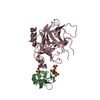
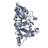
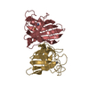
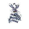
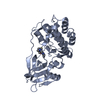
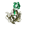
The biological assembly is the macromolecular complex in the asymmetric unit and consists of two DNA binding domains (EcR and RXR), bound to an ecdysone response element (IR-1)
-
Components
-
DNA chain , 2 types, 2 molecules CD
#1: DNA chain
EcdsyoneResponseElement
Mass: 5501.567 Da / Num. of mol.: 1 / Source method: obtained synthetically
#2: DNA chain
EcdysoneResponseElement
Mass: 5492.553 Da / Num. of mol.: 1 / Source method: obtained synthetically
-
Protein , 2 types, 2 molecules AB
#3: Protein
RetinoicacidreceptorRXR-alpha
Mass: 9375.969 Da / Num. of mol.: 1 / Fragment: Retinoid X Receptor DNA binding domain Source method: isolated from a genetically manipulated source Source: (gene. exp.) Homo sapiens (human) / Gene: RXRA / Plasmid: pGEX-4T1 / Species (production host): Escherichia coli / Production host: Escherichia coli BL21(DE3) (bacteria) / Strain (production host): BL21DE3 / References: UniProt: P19793
In the structure databanks used in Yorodumi, some data are registered as the other names, "COVID-19 virus" and "2019-nCoV". Here are the details of the virus and the list of structure data.
Jan 31, 2019. EMDB accession codes are about to change! (news from PDBe EMDB page)
EMDB accession codes are about to change! (news from PDBe EMDB page)
The allocation of 4 digits for EMDB accession codes will soon come to an end. Whilst these codes will remain in use, new EMDB accession codes will include an additional digit and will expand incrementally as the available range of codes is exhausted. The current 4-digit format prefixed with “EMD-” (i.e. EMD-XXXX) will advance to a 5-digit format (i.e. EMD-XXXXX), and so on. It is currently estimated that the 4-digit codes will be depleted around Spring 2019, at which point the 5-digit format will come into force.
The EM Navigator/Yorodumi systems omit the EMD- prefix.
Related info.:Q: What is EMD? / ID/Accession-code notation in Yorodumi/EM Navigator
Yorodumi is a browser for structure data from EMDB, PDB, SASBDB, etc.
This page is also the successor to EM Navigator detail page, and also detail information page/front-end page for Omokage search.
The word "yorodu" (or yorozu) is an old Japanese word meaning "ten thousand". "mi" (miru) is to see.
Related info.:EMDB / PDB / SASBDB / Comparison of 3 databanks / Yorodumi Search / Aug 31, 2016. New EM Navigator & Yorodumi / Yorodumi Papers / Jmol/JSmol / Function and homology information / Changes in new EM Navigator and Yorodumi
 Movie
Movie Controller

 Yorodumi
 Open data
 Basic information
 Components
 Keywords
 Ecdysone receptor /
  Function and homology information

 Authors
 Citation
 Structure visualization
 Downloads & links
 Other downloads
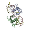
 PDBj
PDBj


 Assembly
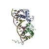


 Mass: 65.409 Da / Num. of mol.: 4 / Source method: obtained synthetically / Formula: Zn
 Sample preparation
 / Beamline: 19-ID / Wavelength: 1.2834, 1.2837, 1.2155
 Processing