 Movie
Movie Controller


+ Open data

- Basic information
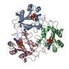
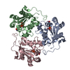
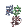
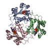
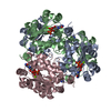
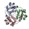
| Entry | Database: PDB / ID: 1hiy | ||||||
|---|---|---|---|---|---|---|---|
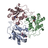
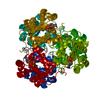
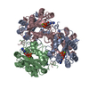
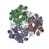
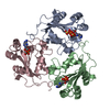
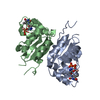
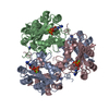
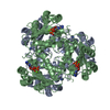
| Title | Binding of nucleotides to NDP kinase | ||||||
 Components Components | NUCLEOSIDE DIPHOSPHATE KINASE Nucleoside-diphosphate kinase Nucleoside-diphosphate kinase | ||||||
 Keywords Keywords | TRANSFERASE / METABOLIC ROLE / KINASE | ||||||
| Function / homology |  Function and homology information Function and homology informationdGTP biosynthetic process from dGDP / Azathioprine ADME / Ribavirin ADME / asexual reproduction / Interconversion of nucleotide di- and triphosphates / Neutrophil degranulation / nucleoside triphosphate biosynthetic process / negative regulation of pinocytosis / nucleoside-diphosphate kinase / UTP biosynthetic process ...dGTP biosynthetic process from dGDP / Azathioprine ADME / Ribavirin ADME / asexual reproduction / Interconversion of nucleotide di- and triphosphates / Neutrophil degranulation / nucleoside triphosphate biosynthetic process / negative regulation of pinocytosis / nucleoside-diphosphate kinase / UTP biosynthetic process / CTP biosynthetic process / negative regulation of exocytosis / negative regulation of phagocytosis / GTP biosynthetic process / nucleoside diphosphate kinase activity / translational elongation / phagocytic vesicle / secretory granule / response to bacterium / actin cytoskeleton organization / cytoskeleton / ribosome / G protein-coupled receptor signaling pathway / phosphorylation / ATP binding / metal ion binding / plasma membrane / cytoplasmSimilarity search - Function | ||||||
| Biological species |  DICTYOSTELIUM DISCOIDEUM (eukaryote) DICTYOSTELIUM DISCOIDEUM (eukaryote) | ||||||
| Method | X-RAY DIFFRACTION / SYNCHROTRON / MOLECULAR REPLACEMENT / Resolution: 2.6 Å | ||||||
 Authors Authors | Cervoni, L. / Lascu, I. / Xu, Y. / Gonin, P. / Morr, M. / Merouani, M. / Janin, J. / Giartoso, A. | ||||||
 Citation Citation | Journal: Biochemistry / Year: 2001 Title: Binding of Nucleotides to Nucleoside Diphosphate Kinase: A Calorimetric Study. Authors: Cervoni, L. / Lascu, I. / Xu, Y. / Gonin, P. / Morr, M. / Merouani, M. / Janin, J. / Giartosio, A. | ||||||
| History |
|
- Structure visualization
| Structure viewer | Molecule: MolmilJmol/JSmol |
|---|
- Downloads & links
-Download
| PDBx/mmCIF format | 1hiy.cif.gz | 100.6 KB | Display | PDBx/mmCIF format |
|---|---|---|---|---|
| PDB format | pdb1hiy.ent.gz | 77.2 KB | Display | PDB format |
| PDBx/mmJSON format | 1hiy.json.gz | Tree view | PDBx/mmJSON format | |
| Others |  Other downloads Other downloads |
-Validation report
| Arichive directory | https://data.pdbj.org/pub/pdb/validation_reports/hi/1hiyftp://data.pdbj.org/pub/pdb/validation_reports/hi/1hiy | HTTPS FTP |
|---|
-Related structure data
| Related structure data |  1kdnS S: Starting model for refinement |
|---|---|
| Similar structure data |


-Links
 PDBj
PDBj
- Assembly
| Deposited unit | 
| ||||||||
|---|---|---|---|---|---|---|---|---|---|
| 1 | 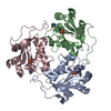
| ||||||||
| Unit cell |
|
-Components
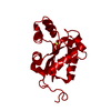
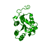
| #1: Protein | Nucleoside-diphosphate kinase Mass: 16816.336 Da / Num. of mol.: 3 Source method: isolated from a genetically manipulated source Details: 3'AMINO-ADP / Source: (gene. exp.) DICTYOSTELIUM DISCOIDEUM (eukaryote) / Production host:  ESCHERICHIA COLI (E. coli) / References: UniProt: P22887, nucleoside-diphosphate kinase ESCHERICHIA COLI (E. coli) / References: UniProt: P22887, nucleoside-diphosphate kinase#2: Chemical |   Mass: 426.216 Da / Num. of mol.: 3 / Source method: obtained synthetically / Formula: C10H16N6O9P2 Mass: 426.216 Da / Num. of mol.: 3 / Source method: obtained synthetically / Formula: C10H16N6O9P2#3: Water | ChemComp-HOH / | Water Mass: 18.015 Da / Num. of mol.: 41 / Source method: isolated from a natural source / Formula: H2O Mass: 18.015 Da / Num. of mol.: 41 / Source method: isolated from a natural source / Formula: H2OCompound details | PLAYS A MAJOR ROLE IN THE SYNTHESIS OF NUCLEOSIDE | |
|---|
-Experimental details
-Experiment
| Experiment | Method: X-RAY DIFFRACTION / Number of used crystals: 1 |
|---|
- Sample preparation
| Crystal | Density Matthews: 2.26 Å3/Da / Density % sol: 31 % | ||||||||||||||||||||||||||||||||||||
|---|---|---|---|---|---|---|---|---|---|---|---|---|---|---|---|---|---|---|---|---|---|---|---|---|---|---|---|---|---|---|---|---|---|---|---|---|---|
| Crystal grow | Method: vapor diffusion, hanging drop / pH: 8.5 Details: METHOD: HANGING DROP IN DROP: 5MG/ML PROTEIN, 50 MM TRIS HCL PH8.5, 8.5MM 3'-AMINO-ADP,15-16% PEG550, 20MM MGCL2 IN WELL: 30-32% PEG550, 50MM TRISHCL PH8.5., pH 8.50 | ||||||||||||||||||||||||||||||||||||
| Crystal grow | *PLUS pH: 8 / Method: vapor diffusion, hanging drop | ||||||||||||||||||||||||||||||||||||
| Components of the solutions | *PLUS
|
-Data collection
| Diffraction | Mean temperature: 277 K |
|---|---|
| Diffraction source | Source: SYNCHROTRON / Site: LURE  / Beamline: DW32 / Wavelength: 0.97 / Wavelength: 0.97 Å / Beamline: DW32 / Wavelength: 0.97 / Wavelength: 0.97 Å |
| Detector | Detector: IMAGE PLATE |
| Radiation | Protocol: SINGLE WAVELENGTH / Monochromatic (M) / Laue (L): M / Scattering type: x-ray |
| Radiation wavelength | Wavelength: 0.97 Å / Relative weight: 1 |
| Reflection | Resolution: 2.2→15 Å / Num. obs: 23760 / % possible obs: 99.7 % / Observed criterion σ(I): 2 / Redundancy: 4 % / Biso Wilson estimate: 27 Å2 / Rmerge(I) obs: 0.049 |
| Reflection | *PLUS Lowest resolution: 15 Å |
- Processing
| Software |
| ||||||||||||||||||||||||||||||||||||||||||||||||||||||||||||
|---|---|---|---|---|---|---|---|---|---|---|---|---|---|---|---|---|---|---|---|---|---|---|---|---|---|---|---|---|---|---|---|---|---|---|---|---|---|---|---|---|---|---|---|---|---|---|---|---|---|---|---|---|---|---|---|---|---|---|---|---|---|
| Refinement | Method to determine structure: MOLECULAR REPLACEMENT Starting model: 1KDN Resolution: 2.6→15 Å / σ(F): 2
| ||||||||||||||||||||||||||||||||||||||||||||||||||||||||||||
| Displacement parameters | Biso mean: 30.3 Å2 | ||||||||||||||||||||||||||||||||||||||||||||||||||||||||||||
| Refinement step | Cycle: LAST / Resolution: 2.6→15 Å
| ||||||||||||||||||||||||||||||||||||||||||||||||||||||||||||
| Refine LS restraints |
| ||||||||||||||||||||||||||||||||||||||||||||||||||||||||||||
| Refine LS restraints NCS | NCS model details: BACKBOND RESTRAINTS | ||||||||||||||||||||||||||||||||||||||||||||||||||||||||||||
| Software | *PLUS Name: X-PLOR / Version: 3.8 / Classification: refinement | ||||||||||||||||||||||||||||||||||||||||||||||||||||||||||||
| Refinement | *PLUS Lowest resolution: 15 Å / σ(F): 2 | ||||||||||||||||||||||||||||||||||||||||||||||||||||||||||||
| Solvent computation | *PLUS | ||||||||||||||||||||||||||||||||||||||||||||||||||||||||||||
| Displacement parameters | *PLUS | ||||||||||||||||||||||||||||||||||||||||||||||||||||||||||||
| Refine LS restraints | *PLUS
|
