 Movie
Movie Controller

[English] 日本語
 Yorodumi
- PDB-1ndc: X-RAY STRUCTURE OF NUCLEOSIDE DIPHOSPHATE KINASE COMPLEXED WITH D... -
+ Open data

- Basic information
| Entry | Database: PDB / ID: 1ndc | |||||||||
|---|---|---|---|---|---|---|---|---|---|---|
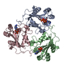
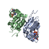
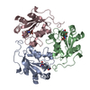
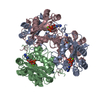
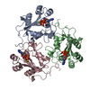
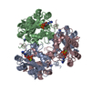
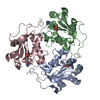
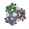
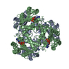
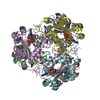
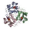
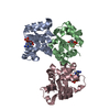
| Title | X-RAY STRUCTURE OF NUCLEOSIDE DIPHOSPHATE KINASE COMPLEXED WITH DTDP AND MG2+ AT 2 A RESOLUTION | |||||||||
 Components Components | NUCLEOSIDE DIPHOSPHATE KINASE | |||||||||
 Keywords Keywords | PHOSPHOTRANSFERASE / NUCLEOSIDE TRIPHOSPHATE / NUCLEOSIDE DIPHOSPHATE | |||||||||
| Function / homology |  Function and homology information Function and homology information: / Azathioprine ADME / Ribavirin ADME / asexual reproduction / Interconversion of nucleotide di- and triphosphates / Neutrophil degranulation / negative regulation of pinocytosis / nucleoside triphosphate biosynthetic process / nucleoside-diphosphate kinase / UTP biosynthetic process ...: / Azathioprine ADME / Ribavirin ADME / asexual reproduction / Interconversion of nucleotide di- and triphosphates / Neutrophil degranulation / negative regulation of pinocytosis / nucleoside triphosphate biosynthetic process / nucleoside-diphosphate kinase / UTP biosynthetic process / CTP biosynthetic process / nucleoside diphosphate kinase activity / GTP biosynthetic process / negative regulation of exocytosis / negative regulation of phagocytosis / translational elongation / phagocytic vesicle / secretory granule / response to bacterium / actin cytoskeleton organization / cytoskeleton / ribosome / G protein-coupled receptor signaling pathway / ATP binding / metal ion binding / plasma membrane / cytoplasm Similarity search - Function | |||||||||
| Biological species |  | |||||||||
| Method |  X-RAY DIFFRACTION / Resolution: 2 Å X-RAY DIFFRACTION / Resolution: 2 Å | |||||||||
 Authors Authors | Cherfils, J. / Morera, S. / Janin, J. | |||||||||
 Citation Citation | Journal: Biochemistry / Year: 1994 Title: X-ray structure of nucleoside diphosphate kinase complexed with thymidine diphosphate and Mg2+ at 2-A resolution. Authors: Cherfils, J. / Morera, S. / Lascu, I. / Veron, M. / Janin, J. #1: Journal: Biochemistry / Year: 1994Title: Adp Binding and the Active Site of Nucleoside Diphosphate Kinase Authors: Morera, S. / Lascu, I. / Dumas, C. / Lebras, G. / Briozzo, P. / Veron, M. / Janin, J. #2: Journal: Embo J. / Year: 1992Title: X-Ray Structure of Nucleoside Diphosphate Kinase Authors: Dumas, C. / Lascu, I. / Morera, S. / Glaser, P. / Fourme, R. / Wallet, V. / Lacombe, M.L. / Veron, M. / Janin, J. | |||||||||
| History |
|
- Structure visualization
| Structure viewer | Molecule: MolmilJmol/JSmol |
|---|
- Downloads & links
-Download
| PDBx/mmCIF format | 1ndc.cif.gz | 44.4 KB | Display | PDBx/mmCIF format |
|---|---|---|---|---|
| PDB format | pdb1ndc.ent.gz | 30.5 KB | Display | PDB format |
| PDBx/mmJSON format | 1ndc.json.gz | Tree view | PDBx/mmJSON format | |
| Others |  Other downloads Other downloads |
-Validation report
| Arichive directory | https://data.pdbj.org/pub/pdb/validation_reports/nd/1ndcftp://data.pdbj.org/pub/pdb/validation_reports/nd/1ndc | HTTPS FTP |
|---|
-Related structure data
| Similar structure data |
|---|
-Links
 PDBj
PDBj
- Assembly
| Deposited unit | 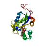
| ||||||||
|---|---|---|---|---|---|---|---|---|---|
| 1 | x 6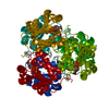
| ||||||||
| Unit cell |
| ||||||||
| Components on special symmetry positions |
|
-Components
| #1: Protein | Mass: 16816.336 Da / Num. of mol.: 1 Source method: isolated from a genetically manipulated source Source: (gene. exp.) |
|---|---|
| #2: Chemical | ChemComp-MG /   Mass: 24.305 Da / Num. of mol.: 1 / Source method: obtained synthetically / Formula: Mg Mass: 24.305 Da / Num. of mol.: 1 / Source method: obtained synthetically / Formula: Mg |
| #3: Chemical | ChemComp-TYD /   Mass: 402.188 Da / Num. of mol.: 1 / Source method: obtained synthetically / Formula: C10H16N2O11P2 Mass: 402.188 Da / Num. of mol.: 1 / Source method: obtained synthetically / Formula: C10H16N2O11P2 |
| #4: Water | ChemComp-HOH /  Mass: 18.015 Da / Num. of mol.: 105 / Source method: isolated from a natural source / Formula: H2O Mass: 18.015 Da / Num. of mol.: 105 / Source method: isolated from a natural source / Formula: H2O |
| Nonpolymer details | A MG++ ION IS ASSOCIATED |
-Experimental details
-Experiment
| Experiment | Method: X-RAY DIFFRACTION |
|---|
- Sample preparation
| Crystal | Density Matthews: 2.92 Å3/Da / Density % sol: 57.94 % | |||||||||||||||||||||||||||||||||||||||||||||||||
|---|---|---|---|---|---|---|---|---|---|---|---|---|---|---|---|---|---|---|---|---|---|---|---|---|---|---|---|---|---|---|---|---|---|---|---|---|---|---|---|---|---|---|---|---|---|---|---|---|---|---|
| Crystal grow | *PLUS Temperature: 18 ℃ / Method: vapor diffusion, hanging drop | |||||||||||||||||||||||||||||||||||||||||||||||||
| Components of the solutions | *PLUS
|
-Data collection
| Radiation | Scattering type: x-ray |
|---|---|
| Radiation wavelength | Relative weight: 1 |
| Reflection | *PLUS Highest resolution: 2 Å / Lowest resolution: 16 Å / Num. obs: 13562 / % possible obs: 99.6 % / Num. measured all: 71549 / Rmerge(I) obs: 0.068 |
- Processing
| Software |
| ||||||||||||||||||||||||||||||||||||||||||||||||||||||||||||
|---|---|---|---|---|---|---|---|---|---|---|---|---|---|---|---|---|---|---|---|---|---|---|---|---|---|---|---|---|---|---|---|---|---|---|---|---|---|---|---|---|---|---|---|---|---|---|---|---|---|---|---|---|---|---|---|---|---|---|---|---|---|
| Refinement | Resolution: 2→16 Å / σ(F): 0 /
| ||||||||||||||||||||||||||||||||||||||||||||||||||||||||||||
| Refinement step | Cycle: LAST / Resolution: 2→16 Å
| ||||||||||||||||||||||||||||||||||||||||||||||||||||||||||||
| Refine LS restraints |
| ||||||||||||||||||||||||||||||||||||||||||||||||||||||||||||
| Software | *PLUS Name: X-PLOR / Classification: refinement | ||||||||||||||||||||||||||||||||||||||||||||||||||||||||||||
| Refinement | *PLUS Num. reflection all: 13562 / Rfactor all: 0.183 / Rfactor Rwork: 0.183 | ||||||||||||||||||||||||||||||||||||||||||||||||||||||||||||
| Solvent computation | *PLUS | ||||||||||||||||||||||||||||||||||||||||||||||||||||||||||||
| Displacement parameters | *PLUS Biso mean: 11.9 Å2 | ||||||||||||||||||||||||||||||||||||||||||||||||||||||||||||
| Refine LS restraints | *PLUS Type: x_angle_d / Dev ideal: 1.6 |
