 Movie
Movie Controller

[English] 日本語
 Yorodumi
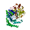
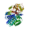
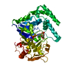
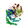
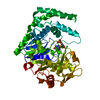
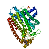
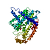
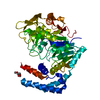
- PDB-1cib: STRUCTURE OF ADENYLOSUCCINATE SYNTHETASE FROM E. COLI COMPLEXED W... -
+ Open data

- Basic information
| Entry | Database: PDB / ID: 1cib | ||||||
|---|---|---|---|---|---|---|---|
| Title | STRUCTURE OF ADENYLOSUCCINATE SYNTHETASE FROM E. COLI COMPLEXED WITH GDP, IMP, HADACIDIN, AND NO3 | ||||||
 Components Components | ADENYLOSUCCINATE SYNTHETASE Adenylosuccinate synthase Adenylosuccinate synthase | ||||||
 Keywords Keywords | LIGASE / GTP-HYDROLYSING ENZYMES / PURINE 2 NUCLEOTIDE BIOSYNTHESIS / GDP | ||||||
| Function / homology |  Function and homology informationadenylosuccinate synthase / adenylosuccinate synthase activity / adenosine biosynthetic process / IMP metabolic process / 'de novo' AMP biosynthetic process / nucleobase-containing small molecule interconversion / purine nucleotide biosynthetic process / guanosine tetraphosphate binding / DNA damage response / GTP binding ...adenylosuccinate synthase / adenylosuccinate synthase activity / adenosine biosynthetic process / IMP metabolic process / 'de novo' AMP biosynthetic process / nucleobase-containing small molecule interconversion / purine nucleotide biosynthetic process / guanosine tetraphosphate binding / DNA damage response / GTP binding / magnesium ion binding / membrane / cytosol / cytoplasm Function and homology informationadenylosuccinate synthase / adenylosuccinate synthase activity / adenosine biosynthetic process / IMP metabolic process / 'de novo' AMP biosynthetic process / nucleobase-containing small molecule interconversion / purine nucleotide biosynthetic process / guanosine tetraphosphate binding / DNA damage response / GTP binding ...adenylosuccinate synthase / adenylosuccinate synthase activity / adenosine biosynthetic process / IMP metabolic process / 'de novo' AMP biosynthetic process / nucleobase-containing small molecule interconversion / purine nucleotide biosynthetic process / guanosine tetraphosphate binding / DNA damage response / GTP binding / magnesium ion binding / membrane / cytosol / cytoplasmSimilarity search - Function | ||||||
| Biological species |  Escherichia coli (E. coli) Escherichia coli (E. coli) | ||||||
| Method | X-RAY DIFFRACTION / MOLECULAR REPLACEMENT / Resolution: 2.3 Å | ||||||
 Authors Authors | Hou, Z. / Cashel, M. / Fromm, H.J. / Honzatko, R.B. | ||||||
 Citation Citation | Journal: J.Biol.Chem. / Year: 1999 Title: Effectors of the stringent response target the active site of Escherichia coli adenylosuccinate synthetase. Authors: Hou, Z. / Cashel, M. / Fromm, H.J. / Honzatko, R.B. | ||||||
| History |
|
- Structure visualization
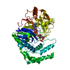
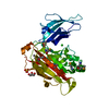
| Structure viewer | Molecule: MolmilJmol/JSmol |
|---|
- Downloads & links
-Download
| PDBx/mmCIF format | 1cib.cif.gz | 108.9 KB | Display | PDBx/mmCIF format |
|---|---|---|---|---|
| PDB format | pdb1cib.ent.gz | 80.9 KB | Display | PDB format |
| PDBx/mmJSON format | 1cib.json.gz | Tree view | PDBx/mmJSON format | |
| Others |  Other downloads Other downloads |
-Validation report
| Arichive directory | https://data.pdbj.org/pub/pdb/validation_reports/ci/1cibftp://data.pdbj.org/pub/pdb/validation_reports/ci/1cib | HTTPS FTP |
|---|
-Related structure data
| Related structure data | 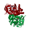 1ch8C 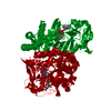 1gimS S: Starting model for refinement C: citing same article ( |
|---|---|
| Similar structure data |
-Links
 PDBj
PDBj- Assembly
| Deposited unit | 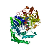
| ||||||||
|---|---|---|---|---|---|---|---|---|---|
| 1 |
| ||||||||
| 2 | 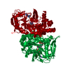
| ||||||||
| Unit cell |
|
-Components
-Protein , 1 types, 1 molecules A
| #1: Protein | Adenylosuccinate synthase / AMPSASE Mass: 47269.598 Da / Num. of mol.: 1 Source method: isolated from a genetically manipulated source Source: (gene. exp.) Escherichia coli (E. coli) / Species: coli / Strain: K-12 / Plasmid: PTRC99A / Production host: Escherichia coli (E. coli) / Strain (production host): PUR A H1238 / References: UniProt: P0A7D4, adenylosuccinate synthase |
|---|
-Non-polymers , 6 types, 415 molecules 


| #2: Chemical | ChemComp-NO3 / Nitrate Mass: 62.005 Da / Num. of mol.: 1 / Source method: obtained synthetically / Formula: NO3 Mass: 62.005 Da / Num. of mol.: 1 / Source method: obtained synthetically / Formula: NO3 |
|---|---|
| #3: Chemical | ChemComp-MG /  Mass: 24.305 Da / Num. of mol.: 1 / Source method: obtained synthetically / Formula: Mg Mass: 24.305 Da / Num. of mol.: 1 / Source method: obtained synthetically / Formula: Mg |
| #4: Chemical | ChemComp-GDP / Guanosine diphosphate Type: RNA linking / Mass: 443.201 Da / Num. of mol.: 1 / Source method: obtained synthetically / Formula: C10H15N5O11P2 / Comment: GDP, energy-carrying molecule*YM Type: RNA linking / Mass: 443.201 Da / Num. of mol.: 1 / Source method: obtained synthetically / Formula: C10H15N5O11P2 / Comment: GDP, energy-carrying molecule*YM |
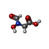
| #5: Chemical | ChemComp-HDA / Hadacidin Mass: 119.076 Da / Num. of mol.: 1 / Source method: obtained synthetically / Formula: C3H5NO4 / Comment: anticancer, medication*YM Mass: 119.076 Da / Num. of mol.: 1 / Source method: obtained synthetically / Formula: C3H5NO4 / Comment: anticancer, medication*YM |
| #6: Chemical | ChemComp-IMP / Inosinic acid Mass: 348.206 Da / Num. of mol.: 1 / Source method: obtained synthetically / Formula: C10H13N4O8P Mass: 348.206 Da / Num. of mol.: 1 / Source method: obtained synthetically / Formula: C10H13N4O8P |
| #7: Water | ChemComp-HOH / WaterMass: 18.015 Da / Num. of mol.: 410 / Source method: isolated from a natural source / Formula: H2O |
-Experimental details
-Experiment
| Experiment | Method: X-RAY DIFFRACTION / Number of used crystals: 1 |
|---|
- Sample preparation
| Crystal | Density Matthews: 3.14 Å3/Da / Density % sol: 60.86 % | ||||||||||||||||||||||||||||||||||||||||||||||||||||||
|---|---|---|---|---|---|---|---|---|---|---|---|---|---|---|---|---|---|---|---|---|---|---|---|---|---|---|---|---|---|---|---|---|---|---|---|---|---|---|---|---|---|---|---|---|---|---|---|---|---|---|---|---|---|---|---|
| Crystal grow | pH: 6.5 / Details: 16% PEG 8000, 100MM NA-CACODYLATE (PH 6.5) | ||||||||||||||||||||||||||||||||||||||||||||||||||||||
| Crystal grow | *PLUS Method: vapor diffusion, hanging drop | ||||||||||||||||||||||||||||||||||||||||||||||||||||||
| Components of the solutions | *PLUS
|
-Data collection
| Diffraction | Mean temperature: 120 K |
|---|---|
| Diffraction source | Source: ROTATING ANODE / Type: SIEMENS / Wavelength: 1.5418 |
| Detector | Type: SIEMENS / Detector: AREA DETECTOR / Date: Feb 1, 1997 |
| Radiation | Protocol: SINGLE WAVELENGTH / Monochromatic (M) / Laue (L): M / Scattering type: x-ray |
| Radiation wavelength | Wavelength: 1.5418 Å / Relative weight: 1 |
| Reflection | Resolution: 2.2→50 Å / Num. obs: 29374 / % possible obs: 99 % / Redundancy: 2.5 % / Rmerge(I) obs: 0.085 |
| Reflection shell | Resolution: 2.2→2.4 Å / % possible all: 97 |
| Reflection | *PLUS % possible obs: 97 % / Num. measured all: 154316 |
| Reflection shell | *PLUS % possible obs: 93 % |
- Processing
| Software |
| ||||||||||||||||||||||||||||||||||||||||||||||||||||||||||||
|---|---|---|---|---|---|---|---|---|---|---|---|---|---|---|---|---|---|---|---|---|---|---|---|---|---|---|---|---|---|---|---|---|---|---|---|---|---|---|---|---|---|---|---|---|---|---|---|---|---|---|---|---|---|---|---|---|---|---|---|---|---|
| Refinement | Method to determine structure: MOLECULAR REPLACEMENT Starting model: 1GIM Resolution: 2.3→6 Å / Rfactor Rfree error: 0.005 / Data cutoff low absF: 0 / Cross valid method: THROUGHOUT / σ(F): 0
| ||||||||||||||||||||||||||||||||||||||||||||||||||||||||||||
| Displacement parameters | Biso mean: 25 Å2 | ||||||||||||||||||||||||||||||||||||||||||||||||||||||||||||
| Refinement step | Cycle: LAST / Resolution: 2.3→6 Å
| ||||||||||||||||||||||||||||||||||||||||||||||||||||||||||||
| Refine LS restraints |
| ||||||||||||||||||||||||||||||||||||||||||||||||||||||||||||
| Xplor file |
| ||||||||||||||||||||||||||||||||||||||||||||||||||||||||||||
| Software | *PLUS Name: X-PLOR / Version: 3.1 / Classification: refinement | ||||||||||||||||||||||||||||||||||||||||||||||||||||||||||||
| Refinement | *PLUS Highest resolution: 2.3 Å / Lowest resolution: 6 Å / σ(F): 0 / % reflection Rfree: 10 % / Rfactor obs: 0.17 / Rfactor Rfree: 0.24 / Rfactor Rwork: 0.17 | ||||||||||||||||||||||||||||||||||||||||||||||||||||||||||||
| Solvent computation | *PLUS | ||||||||||||||||||||||||||||||||||||||||||||||||||||||||||||
| Displacement parameters | *PLUS Biso mean: 25 Å2 |
