Mass: 18.015 Da / Num. of mol.: 166 / Source method: isolated from a natural source / Formula: H2O
-
Details
Has protein modification
Y
Sequence details
THIS CONSTRUCT (RESIDUES 21-375) WAS EXPRESSED WITH A PURIFICATION TAG MGSDKIHHHHHHENLYFQG. THE TAG ...THIS CONSTRUCT (RESIDUES 21-375) WAS EXPRESSED WITH A PURIFICATION TAG MGSDKIHHHHHHENLYFQG. THE TAG WAS REMOVED WITH TEV PROTEASE LEAVING ONLY A GLYCINE (0) FOLLOWED BY THE TARGET SEQUENCE.
-
Experimental details
-
Experiment
Experiment
Method: X-RAY DIFFRACTION / Number of used crystals: 1
-
Sample preparation
Crystal
Density Matthews: 2.76 Å3/Da / Density % sol: 55.43 %
Crystal grow
Temperature: 277 K / Method: vapor diffusion, sitting drop / pH: 9 Details: 0.1M bicine pH 9, 2.4M ammonium sulfate, NANODROP, VAPOR DIFFUSION, SITTING DROP, temperature 277K
Type: DECTRIS PILATUS 6M / Detector: PIXEL / Date: Jan 24, 2013 Details: Flat mirror (vertical focusing); single crystal Si(111) bent monochromator (horizontal focusing)
Radiation
Monochromator: single crystal Si(111) bent / Protocol: MAD / Monochromatic (M) / Laue (L): M / Scattering type: x-ray
Radiation wavelength
ID
Wavelength (Å)
Relative weight
1
0.91837
1
2
0.97932
1
Reflection
Resolution: 2.1→28.711 Å / Num. obs: 25691 / % possible obs: 99.5 % / Observed criterion σ(I): -3 / Biso Wilson estimate: 33.623 Å2 / Rmerge(I) obs: 0.106 / Net I/σ(I): 10.21
Reflection shell
Diffraction-ID: 1
Resolution (Å)
Highest resolution (Å)
Rmerge(I) obs
Mean I/σ(I) obs
Num. measured obs
Num. unique obs
% possible all
2.1-2.17
0.862
1.6
16286
4605
99
2.17-2.26
0.7
2
18322
5171
99.7
2.26-2.36
0.614
2.3
16849
4826
99.5
2.36-2.49
0.496
2.8
17367
5175
99.4
2.49-2.64
0.323
4
16228
4766
99.6
2.64-2.85
0.241
5.6
18434
5161
99.6
2.85-3.13
0.141
9.2
17070
4843
99.6
3.13-3.59
0.073
16.1
17182
5032
99.4
3.59-4.51
0.042
26.8
17508
4913
99.7
4.51
0.033
31.4
17326
5011
99.1
-
Phasing
Phasing
Method: MAD
-
Processing
Software
Name
Version
Classification
NB
MolProbity
3beta29
modelbuilding
PDB_EXTRACT
3.1
dataextraction
SHELX
phasing
SHARP
phasing
XSCALE
July4, 2012
datascaling
REFMAC
5.7.0032
refinement
XDS
datareduction
SHELXD
phasing
autoSHARP
phasing
Refinement
Method to determine structure: MAD / Resolution: 2.1→28.711 Å / Cor.coef. Fo:Fc: 0.963 / Cor.coef. Fo:Fc free: 0.94 / Occupancy max: 1 / Occupancy min: 0.35 / SU B: 8.247 / SU ML: 0.11 / Cross valid method: THROUGHOUT / σ(F): 0 / ESU R: 0.176 / ESU R Free: 0.163 Stereochemistry target values: MAXIMUM LIKELIHOOD WITH PHASES Details: 1. HYDROGENS HAVE BEEN ADDED IN THE RIDING POSITIONS. 2 .A MET-INHIBITION PROTOCOL WAS USED FOR SELENOMETHIONINE INCORPORATION DURING PROTEIN EXPRESSION. THE OCCUPANCY OF THE SE ATOMS IN THE ...Details: 1. HYDROGENS HAVE BEEN ADDED IN THE RIDING POSITIONS. 2 .A MET-INHIBITION PROTOCOL WAS USED FOR SELENOMETHIONINE INCORPORATION DURING PROTEIN EXPRESSION. THE OCCUPANCY OF THE SE ATOMS IN THE MSE RESIDUES WAS REDUCED TO 0.75 FOR THE REDUCED SCATTERING POWER DUE TO PARTIAL S-MET INCORPORATION. 3. SODIUM (NA), CHLORIDE (CL), AND GLYCEROL (GOL) FROM THE CRYSTALLIZATION/CRYO CONDITIONS HAVE BEEN MODELED INTO THE STRUCTURE.4.ATOM RECORDS CONTAIN SUM OF TLS AND RESIDUAL B FACTORS. ANISOU RECORD CONTAINS SUM OF TLS AND RESIDUAL U FACTORS. 5.WATERS WERE EXCLUDED FROM AUTOMATIC TLS ASSIGNMENT. 6. AN UNKNOWN ION (UNX) HAS BEEN MODELED BASED ON A PEAK IN THE ANOMALOUS DIFFERENCE FOURIER MAP. THE ION LIKELY CO-PURIFIED WITH THE PROTEIN. X-RAY FLUORESCENCE EXCITATION SPECTRA WERE INCONCLUSIVE IN DETERMINING THE METAL IDENTITY WITH MINOR PEAKS FOR ZN, CU, FE AND CA. FOR THE PURPOSE OF REFINEMENT THE UNX ATOM TYPE X WAS ASSIGNED SCATTERING FACTORS EQUIVALENT TO CA WHICH GAVE A REASONABLE FIT TO THE OBSERVED DENSITY. 7. THE SCATTERING FACTORS FOR SULFUR, CHLORINE, SELENIUM AND THE UNKNOWN X ATOMS WERE ADJUSTED BY REFMAC 5.7.0032 TO ACCOUNT FOR ANOMALOUS DISPERSION BASED ON THE WAVELENGTH 0.91837 A (S F'= 0.16, CL F'= 0.19, SE F'= -1.94, X F'= 0.27). THE CROMER MANN VALUES LISTED IN THE CIF VERSION OF THE FILE INCLUDE THIS CORRECTION.
Rfactor
Num. reflection
% reflection
Selection details
Rfree
0.2202
1307
5.1 %
RANDOM
Rwork
0.1707
-
-
-
obs
0.1731
25691
99.73 %
-
Solvent computation
Ion probe radii: 0.8 Å / Shrinkage radii: 0.8 Å / VDW probe radii: 1.2 Å / Solvent model: BABINET MODEL WITH MASK
In the structure databanks used in Yorodumi, some data are registered as the other names, "COVID-19 virus" and "2019-nCoV". Here are the details of the virus and the list of structure data.
Jan 31, 2019. EMDB accession codes are about to change! (news from PDBe EMDB page)
EMDB accession codes are about to change! (news from PDBe EMDB page)
The allocation of 4 digits for EMDB accession codes will soon come to an end. Whilst these codes will remain in use, new EMDB accession codes will include an additional digit and will expand incrementally as the available range of codes is exhausted. The current 4-digit format prefixed with “EMD-” (i.e. EMD-XXXX) will advance to a 5-digit format (i.e. EMD-XXXXX), and so on. It is currently estimated that the 4-digit codes will be depleted around Spring 2019, at which point the 5-digit format will come into force.
The EM Navigator/Yorodumi systems omit the EMD- prefix.
Related info.:Q: What is EMD? / ID/Accession-code notation in Yorodumi/EM Navigator
Yorodumi is a browser for structure data from EMDB, PDB, SASBDB, etc.
This page is also the successor to EM Navigator detail page, and also detail information page/front-end page for Omokage search.
The word "yorodu" (or yorozu) is an old Japanese word meaning "ten thousand". "mi" (miru) is to see.
Related info.:EMDB / PDB / SASBDB / Comparison of 3 databanks / Yorodumi Search / Aug 31, 2016. New EM Navigator & Yorodumi / Yorodumi Papers / Jmol/JSmol / Function and homology information / Changes in new EM Navigator and Yorodumi
 Movie
Movie Controller

 Yorodumi
 Open data
 Basic information
 Components
 Keywords
 Function and homology information
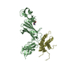
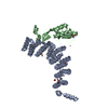
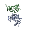
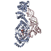
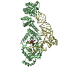
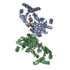
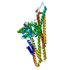
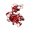
 Bacteroides ovatus (bacteria)
 X-RAY DIFFRACTION /
  Authors
 Citation
 Structure visualization
 Downloads & links
 Other downloads
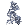

 PDBj
PDBj Assembly
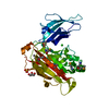


 Num. of mol.: 1 / Source method: obtained synthetically
 Mass: 35.453 Da / Num. of mol.: 1 / Source method: obtained synthetically / Formula: Cl
 Mass: 96.063 Da / Num. of mol.: 1 / Source method: obtained synthetically / Formula: SO4
 Mass: 92.094 Da / Num. of mol.: 4 / Source method: obtained synthetically / Formula: C3H8O3
 Sample preparation
 / Beamline: BL11-1 / Wavelength: 0.91837,0.97932
 Processing