 Movie
Movie Controller

[English] 日本語
 Yorodumi
- PDB-1n78: Crystal structure of Thermus thermophilus glutamyl-tRNA synthetas... -
+ Open data

- Basic information
| Entry | Database: PDB / ID: 1n78 | ||||||
|---|---|---|---|---|---|---|---|
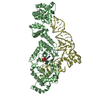
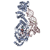
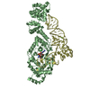
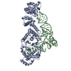
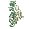
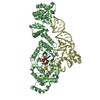
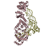
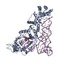
| Title | Crystal structure of Thermus thermophilus glutamyl-tRNA synthetase complexed with tRNA(Glu) and glutamol-AMP. | ||||||
 Components Components |
| ||||||
 Keywords Keywords | ligase/RNA / ERS/tRNA/GOA / RIKEN Structural Genomics/Proteomics Initiative / RSGI / Structural Genomics / ligase-RNA COMPLEX | ||||||
| Function / homology |  Function and homology information Function and homology informationglutamate-tRNA ligase / glutamate-tRNA ligase activity / glutamyl-tRNA aminoacylation / tRNA binding / zinc ion binding / ATP binding / cytosol Similarity search - Function | ||||||
| Biological species |   Thermus thermophilus (bacteria) Thermus thermophilus (bacteria) | ||||||
| Method |  X-RAY DIFFRACTION / SYNCHROTRON / MOLECULAR REPLACEMENT / Resolution: 2.1 Å X-RAY DIFFRACTION / SYNCHROTRON / MOLECULAR REPLACEMENT / Resolution: 2.1 Å | ||||||
 Authors Authors | Sekine, S. / Nureki, O. / Dubois, D.Y. / Bernier, S. / Chenevert, R. / Lapointe, J. / Vassylyev, D.G. / Yokoyama, S. / RIKEN Structural Genomics/Proteomics Initiative (RSGI) | ||||||
 Citation Citation | Journal: EMBO J. / Year: 2003 Title: ATP binding by glutamyl-tRNA synthetase is switched to the productive mode by tRNA binding Authors: Sekine, S. / Nureki, O. / Dubois, D.Y. / Bernier, S. / Chenevert, R. / Lapointe, J. / Vassylyev, D.G. / Yokoyama, S. #1: Journal: Nat.Struct.Biol. / Year: 2001Title: Structural basis for anticodon recognition by discriminating glutamyl-tRNA synthetase Authors: Sekine, S. / Nureki, O. / Shimada, A. / Vassylyev, D.G. / Yokoyama, S. #2: Journal: Science / Year: 1995Title: Architectures of class-defining and specific domains of glutamyl-tRNA synthetase Authors: Nureki, O. / Vassylyev, D.G. / Katayanagi, K. / Shimizu, T. / Sekine, S. / Kigawa, T. / Miyazawa, T. / Yokoyama, S. / Morikawa, K. | ||||||
| History |
|
- Structure visualization
| Structure viewer | Molecule: MolmilJmol/JSmol |
|---|
- Downloads & links
-Download
| PDBx/mmCIF format | 1n78.cif.gz | 300.6 KB | Display | PDBx/mmCIF format |
|---|---|---|---|---|
| PDB format | pdb1n78.ent.gz | 234.2 KB | Display | PDB format |
| PDBx/mmJSON format | 1n78.json.gz | Tree view | PDBx/mmJSON format | |
| Others |  Other downloads Other downloads |
-Validation report
| Arichive directory | https://data.pdbj.org/pub/pdb/validation_reports/n7/1n78ftp://data.pdbj.org/pub/pdb/validation_reports/n7/1n78 | HTTPS FTP |
|---|
-Related structure data
| Related structure data |  1j09C  1n75C  1n77C  1g59S S: Starting model for refinement C: citing same article ( |
|---|---|
| Similar structure data | |
| Other databases |


-Links
 PDBj
PDBj


- Assembly
| Deposited unit | 
| ||||||||
|---|---|---|---|---|---|---|---|---|---|
| 1 | 
| ||||||||
| 2 | 
| ||||||||
| Unit cell |
|
-Components
| #1: RNA chain | Mass: 24105.336 Da / Num. of mol.: 2 / Source method: obtained synthetically Details: This t-RNA occurs from Thermus thermophilus, in vitro transcription #2: Protein | Mass: 53988.727 Da / Num. of mol.: 2 Source method: isolated from a genetically manipulated source Source: (gene. exp.) Thermus thermophilus (bacteria) / Plasmid: pK7 / Species (production host): Escherichia coli / Production host: #3: Chemical |   Mass: 24.305 Da / Num. of mol.: 2 / Source method: obtained synthetically / Formula: Mg Mass: 24.305 Da / Num. of mol.: 2 / Source method: obtained synthetically / Formula: Mg#4: Chemical |   Type: RNA linking / Mass: 461.344 Da / Num. of mol.: 2 / Source method: obtained synthetically / Formula: C15H22N6O9P Type: RNA linking / Mass: 461.344 Da / Num. of mol.: 2 / Source method: obtained synthetically / Formula: C15H22N6O9P#5: Water | ChemComp-HOH / |  Mass: 18.015 Da / Num. of mol.: 751 / Source method: isolated from a natural source / Formula: H2O Mass: 18.015 Da / Num. of mol.: 751 / Source method: isolated from a natural source / Formula: H2O |
|---|
-Experimental details
-Experiment
| Experiment | Method: X-RAY DIFFRACTION / Number of used crystals: 1 |
|---|
- Sample preparation
| Crystal | Density Matthews: 2.52 Å3/Da / Density % sol: 50.81 % | |||||||||||||||||||||||||||||||||||||||||||||||||||||||||||||||
|---|---|---|---|---|---|---|---|---|---|---|---|---|---|---|---|---|---|---|---|---|---|---|---|---|---|---|---|---|---|---|---|---|---|---|---|---|---|---|---|---|---|---|---|---|---|---|---|---|---|---|---|---|---|---|---|---|---|---|---|---|---|---|---|---|
| Crystal grow | Temperature: 293 K / Method: vapor diffusion, hanging drop / pH: 6.7 Details: PEG1500, pH 6.7, VAPOR DIFFUSION, HANGING DROP, temperature 293K | |||||||||||||||||||||||||||||||||||||||||||||||||||||||||||||||
| Components of the solutions |
| |||||||||||||||||||||||||||||||||||||||||||||||||||||||||||||||
| Crystal grow | *PLUS Temperature: 4 or 20 ℃ / pH: 6.5 / Method: vapor diffusion | |||||||||||||||||||||||||||||||||||||||||||||||||||||||||||||||
| Components of the solutions | *PLUS
|
-Data collection
| Diffraction | Mean temperature: 100 K |
|---|---|
| Diffraction source | Source: SYNCHROTRON / Site: SPring-8  / Beamline: BL41XU / Wavelength: 0.9 Å / Beamline: BL41XU / Wavelength: 0.9 Å |
| Detector | Type: MARRESEARCH / Detector: CCD / Date: Jun 12, 2001 |
| Radiation | Monochromator: GRAPHITE / Protocol: SINGLE WAVELENGTH / Monochromatic (M) / Laue (L): M / Scattering type: x-ray |
| Radiation wavelength | Wavelength: 0.9 Å / Relative weight: 1 |
| Reflection | Resolution: 2.1→50 Å / Num. all: 426837 / Num. obs: 92817 / % possible obs: 96.4 % / Observed criterion σ(I): -0.3 / Biso Wilson estimate: 15.5 Å2 / Rmerge(I) obs: 0.074 |
| Reflection shell | Resolution: 2.1→2.2 Å / Rmerge(I) obs: 0.342 / % possible all: 88.5 |
| Reflection | *PLUS Lowest resolution: 50 Å / Num. measured all: 426837 |
| Reflection shell | *PLUS % possible obs: 88.5 % |
- Processing
| Software |
| ||||||||||||||||||||||||||||||||||||
|---|---|---|---|---|---|---|---|---|---|---|---|---|---|---|---|---|---|---|---|---|---|---|---|---|---|---|---|---|---|---|---|---|---|---|---|---|---|
| Refinement | Method to determine structure: MOLECULAR REPLACEMENT Starting model: PDB 1G59 Resolution: 2.1→49.36 Å / Rfactor Rfree error: 0.004 / Data cutoff high absF: 5656722.2 / Data cutoff low absF: 0 / Isotropic thermal model: RESTRAINED / Cross valid method: THROUGHOUT / σ(F): 0
| ||||||||||||||||||||||||||||||||||||
| Solvent computation | Solvent model: FLAT MODEL / Bsol: 52.6101 Å2 / ksol: 0.342137 e/Å3 | ||||||||||||||||||||||||||||||||||||
| Displacement parameters | Biso mean: 35.5 Å2
| ||||||||||||||||||||||||||||||||||||
| Refine analyze |
| ||||||||||||||||||||||||||||||||||||
| Refinement step | Cycle: LAST / Resolution: 2.1→49.36 Å
| ||||||||||||||||||||||||||||||||||||
| Refine LS restraints |
| ||||||||||||||||||||||||||||||||||||
| Refine LS restraints NCS | NCS model details: CONSTR | ||||||||||||||||||||||||||||||||||||
| LS refinement shell | Resolution: 2.1→2.2 Å / Rfactor Rfree error: 0.016 / Total num. of bins used: 8
| ||||||||||||||||||||||||||||||||||||
| Xplor file |
| ||||||||||||||||||||||||||||||||||||
| Refinement | *PLUS Highest resolution: 2.1 Å / Lowest resolution: 50 Å / % reflection Rfree: 5 % | ||||||||||||||||||||||||||||||||||||
| Solvent computation | *PLUS | ||||||||||||||||||||||||||||||||||||
| Displacement parameters | *PLUS | ||||||||||||||||||||||||||||||||||||
| Refine LS restraints | *PLUS
|
