 ムービー
ムービー コントローラー

+ データを開く

- 基本情報
基本情報
| 登録情報 | データベース: SASBDB / ID: SASDF99 |
|---|---|
 試料 試料 | Bovine serum albumin monomer - SEC-SAXS/WAXS coupled to multiangle laser and quasi-elastic light scattering (MALLS and QELS)
|
| 機能・相同性 |  機能・相同性情報 機能・相同性情報cellular response to calcium ion starvation / enterobactin binding / negative regulation of mitochondrial depolarization / toxic substance binding / fatty acid binding / cellular response to starvation / pyridoxal phosphate binding / protein-containing complex / extracellular space / DNA binding ...cellular response to calcium ion starvation / enterobactin binding / negative regulation of mitochondrial depolarization / toxic substance binding / fatty acid binding / cellular response to starvation / pyridoxal phosphate binding / protein-containing complex / extracellular space / DNA binding / extracellular region / metal ion binding / cytoplasm 類似検索 - 分子機能 |
| 生物種 |  |
 登録者 登録者 |
|
- 構造の表示
構造の表示
| 構造ビューア | 分子: MolmilJmol/JSmol |
|---|
- ダウンロードとリンク
 SASDF99
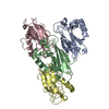


-モデル
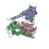
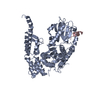
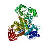
| モデル #3558 |  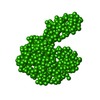 タイプ: dummy / ソフトウェア: (2.3i) / ダミー原子の半径: 1.90 A / 対称性: P1 / カイ2乗値: 1.45 / P-value: 0.298705  Omokage検索でこの集合体の類似形状データを探す (詳細) Omokage検索でこの集合体の類似形状データを探す (詳細) |
|---|---|
| モデル #3560 |   タイプ: atomic / ダミー原子の半径: 1.90 A / カイ2乗値: 2.417 Omokage検索でこの集合体の類似形状データを探す (詳細) |
| モデル #3559 |  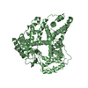 タイプ: atomic / 対称性: P1 / カイ2乗値: 1.548 / P-value: 0.033334 Omokage検索でこの集合体の類似形状データを探す (詳細) |
-試料
| 試料 | 名称: Bovine serum albumin monomer - SEC-SAXS/WAXS coupled to multiangle laser and quasi-elastic light scattering (MALLS and QELS) 試料濃度: 15 mg/ml |
|---|---|
| バッファ | 名称: 50 mM HEPES, 3% v/v glycerol, / pH: 7.5 |
| 要素 #1306 | 名称: BSA / タイプ: protein / 記述: Bovine serum albumin / 分子量: 66.432 / 分子数: 1 / 由来: Bos taurus / 参照: UniProt: P02769 配列: DTHKSEIAHR FKDLGEEHFK GLVLIAFSQY LQQCPFDEHV KLVNELTEFA KTCVADESHA GCEKSLHTLF GDELCKVASL RETYGDMADC CEKQEPERNE CFLSHKDDSP DLPKLKPDPN TLCDEFKADE KKFWGKYLYE IARRHPYFYA PELLYYANKY NGVFQECCQA ...配列: DTHKSEIAHR FKDLGEEHFK GLVLIAFSQY LQQCPFDEHV KLVNELTEFA KTCVADESHA GCEKSLHTLF GDELCKVASL RETYGDMADC CEKQEPERNE CFLSHKDDSP DLPKLKPDPN TLCDEFKADE KKFWGKYLYE IARRHPYFYA PELLYYANKY NGVFQECCQA EDKGACLLPK IETMREKVLA SSARQRLRCA SIQKFGERAL KAWSVARLSQ KFPKAEFVEV TKLVTDLTKV HKECCHGDLL ECADDRADLA KYICDNQDTI SSKLKECCDK PLLEKSHCIA EVEKDAIPEN LPPLTADFAE DKDVCKNYQE AKDAFLGSFL YEYSRRHPEY AVSVLLRLAK EYEATLEECC AKDDPHACYS TVFDKLKHLV DEPQNLIKQN CDQFEKLGEY GFQNALIVRY TRKVPQVSTP TLVEVSRSLG KVGTRCCTKP ESERMPCTED YLSLILNRLC VLHEKTPVSE KVTKCCTESL VNRRPCFSAL TPDETYVPKA FDEKLFTFHA DICTLPDTEK QIKKQTALVE LLKHKPKATE EQLKTVMENF VAFVDKCCAA DDKEACFAVE GPKLVVSTQT ALA |
-実験情報
| ビーム | 設備名称: PETRA III EMBL P12 / 地域: Hamburg / 国: Germany  / 線源: X-ray synchrotron / 波長: 0.124 Å / スペクトロメータ・検出器間距離: 1 mm / 線源: X-ray synchrotron / 波長: 0.124 Å / スペクトロメータ・検出器間距離: 1 mm | ||||||||||||||||||||||||||||||
|---|---|---|---|---|---|---|---|---|---|---|---|---|---|---|---|---|---|---|---|---|---|---|---|---|---|---|---|---|---|---|---|
| 検出器 | 名称: Pilatus 2M | ||||||||||||||||||||||||||||||
| スキャン |  測定日: 2017年4月23日 / 保管温度: 20 °C / セル温度: 20 °C / 照射時間: 1 sec. / フレーム数: 63 / 単位: 1/nm /
| ||||||||||||||||||||||||||||||
| 距離分布関数 P(R) | 
| ||||||||||||||||||||||||||||||
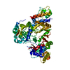
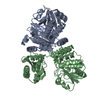
| 結果 |  コメント: Protein powder (Sigma) consisting of BSA monomers, dimers, trimers and higher MW species was dissolved in the SEC-running buffer and then 0.22 micon spin filtered prior to injection ...コメント: Protein powder (Sigma) consisting of BSA monomers, dimers, trimers and higher MW species was dissolved in the SEC-running buffer and then 0.22 micon spin filtered prior to injection onto the SEC column. The sample injection concentration was determined from triplicate UV A280 measurements using an E0.1% of 0.646 (= 1 g/l) calculated from the amino acid sequence (ProtParam). The Rg-correlation through the SEC-SAXS/WAXS peak, the individual unsubtracted SEC-SAXS/WAXS frames as well as the results from coupled MALLS and QELS analysis are included in the full entry zip archive. The quoted experimental molecular weight was determined from the separated monomer peak using MALLS in combination with refractive-index (RI) measurements from the same sample eluting from the column using a split-flow SEC-SAXS/WAXS-light scattering configuration (Graewert et al., (2015) Sci. Reports. 5, 10734: doi: 10.1038/srep10734). The average hydrodynamic radius of the separated monomer was evaluated at 3.4 nm. Two atomistic representations of the protein are displayed: 1) The X-ray crystallography model fit to the SAXS data (PDB:4F5S), middle, and; 2) The same model after normal mode analysis/refinement using SREFLEX (bottom; Panjkovich et al., (2016) Phys. Chem. Chem. Phys. 18(8):5707-5719. doi: 10.1039/c5cp04540a). The complete SREFLEX result summary is included in the full-entry zip archive.
|
