+ データを開く
- 基本情報
基本情報
| 登録情報 | データベース: SASBDB / ID: SASDF94 |
|---|---|
 試料 試料 | Insulin glulisine (Apidra), oligomeric composition
|
 引用 引用 |  日付: 2019 Jul 日付: 2019 Julタイトル: The quaternary structure of insulin glargine and glulisine under formulation conditions 著者: Nagel N / Graewert M / Gao M / Heyse W / Jeffries C / Svergun D |
 登録者 登録者 |
|
- 構造の表示
構造の表示
| 構造ビューア | 分子:  Molmil Molmil Jmol/JSmol Jmol/JSmol |
|---|
- ダウンロードとリンク
-モデル
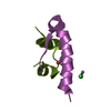
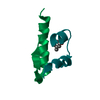
| モデル #2919 | 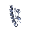 タイプ: atomic / コメント: monomeric unit / カイ2乗値: 1.03 / P-value: 0.574548  Omokage検索でこの集合体の類似形状データを探す (詳細) Omokage検索でこの集合体の類似形状データを探す (詳細) |
|---|---|
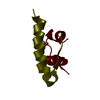
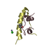
| モデル #2920 | 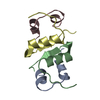 タイプ: atomic / コメント: dimeric unit / カイ2乗値: 1.03 / P-value: 0.574548  Omokage検索でこの集合体の類似形状データを探す (詳細) Omokage検索でこの集合体の類似形状データを探す (詳細) |
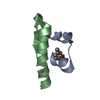
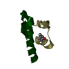
| モデル #2921 | 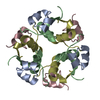 タイプ: atomic / コメント: hexameric unit / カイ2乗値: 1.03 / P-value: 0.574548  Omokage検索でこの集合体の類似形状データを探す (詳細) Omokage検索でこの集合体の類似形状データを探す (詳細) |
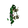
| モデル #2922 | 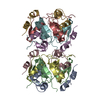 タイプ: atomic / カイ2乗値: 1.03 / P-value: 0.574548  Omokage検索でこの集合体の類似形状データを探す (詳細) Omokage検索でこの集合体の類似形状データを探す (詳細) |
- 試料
試料
 試料 試料 | 名称: Insulin glulisine (Apidra), oligomeric composition / 試料濃度: 3.49 mg/ml |
|---|---|
| バッファ | 名称: Apidra formulation (per ml: 5 mg Sodium chloride, 3.15 mg m-Cresol, 6 mg Trometamol, 0.01 mg Polysorbate 20) pH: 7.3 |
| 要素 #1567 | タイプ: protein / 記述: Insulin glulisine / 分子量: 5.811 / 分子数: 6 配列: GIVEQCCTSI CSLYQLENYC NFVKQHLCGS HLVEALYLVC GERGFFYTPE T |
-実験情報
| ビーム | 設備名称: PETRA III EMBL P12 / 地域: Hamburg / 国: Germany  / 線源: X-ray synchrotron / 波長: 0.1241 Å / スペクトロメータ・検出器間距離: 3.1 mm / 線源: X-ray synchrotron / 波長: 0.1241 Å / スペクトロメータ・検出器間距離: 3.1 mm | |||||||||||||||||||||||||||||
|---|---|---|---|---|---|---|---|---|---|---|---|---|---|---|---|---|---|---|---|---|---|---|---|---|---|---|---|---|---|---|
| 検出器 | 名称: Pilatus 2M | |||||||||||||||||||||||||||||
| スキャン | 測定日: 2017年4月20日 / 保管温度: 20 °C / セル温度: 20 °C / 照射時間: 0.045 sec. / フレーム数: 20 / 単位: 1/A /
| |||||||||||||||||||||||||||||
| 距離分布関数 P(R) |
| |||||||||||||||||||||||||||||
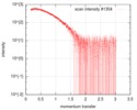
| 結果 | コメント: Here, Apidra measured at 3.49 mg/ml is displayed (formulation concentration). Measurements from 2 dilutions in placebo have been deposited in addition. Mixture analysis of the ...コメント: Here, Apidra measured at 3.49 mg/ml is displayed (formulation concentration). Measurements from 2 dilutions in placebo have been deposited in addition. Mixture analysis of the scattering profiles was performed with the program Oligomer. The SAXS data show that Apidra® primarily consists of hexamers, which dissociate into monomers upon dilution. The presence of dimers was not required to fit the experimental data, and their volume fraction was always essentially zero. A small volume fraction is, however, also made up by larger dodecameric species. The distribution between monomer, dimer, hexamer and dodecamer is documented in the logfile. Calculation of the form factors is based on an internal model (Apidra.pdb) for hexamers and 3W80.pdb for dodecamers.
|
 ムービー
ムービー コントローラー


 SASDF94
