 ムービー
ムービー コントローラー

+ データを開く

- 基本情報
基本情報
| 登録情報 | データベース: SASBDB / ID: SASDF92 |
|---|---|
 試料 試料 | Urokinase plasminogen activator surface receptor, uPAR, T51C-V70C
|
| 機能・相同性 |  機能・相同性情報 機能・相同性情報urokinase plasminogen activator receptor activity / Attachment of GPI anchor to uPAR / positive regulation of homotypic cell-cell adhesion / urokinase plasminogen activator signaling pathway / regulation of plasminogen activation / regulation of fibrinolysis / protein complex involved in cell-matrix adhesion / serine-type endopeptidase complex / Dissolution of Fibrin Clot / extrinsic component of membrane ...urokinase plasminogen activator receptor activity / Attachment of GPI anchor to uPAR / positive regulation of homotypic cell-cell adhesion / urokinase plasminogen activator signaling pathway / regulation of plasminogen activation / regulation of fibrinolysis / protein complex involved in cell-matrix adhesion / serine-type endopeptidase complex / Dissolution of Fibrin Clot / extrinsic component of membrane / positive regulation of DNA binding / positive regulation of epidermal growth factor receptor signaling pathway / negative regulation of intrinsic apoptotic signaling pathway / positive regulation of release of cytochrome c from mitochondria / regulation of proteolysis / regulation of cell adhesion / specific granule membrane / cell projection / positive regulation of protein phosphorylation / chemotaxis / blood coagulation / signaling receptor activity / endoplasmic reticulum lumen / signaling receptor binding / protein domain specific binding / external side of plasma membrane / focal adhesion / Neutrophil degranulation / endoplasmic reticulum membrane / negative regulation of apoptotic process / enzyme binding / cell surface / signal transduction / extracellular region / membrane / plasma membrane 類似検索 - 分子機能 |
| 生物種 |  Homo sapiens (ヒト) Homo sapiens (ヒト) |
 引用 引用 | ジャーナル: J Biol Chem / 年: 2019 タイトル: Did evolution create a flexible ligand-binding cavity in the urokinase receptor through deletion of a plesiotypic disulfide bond? 著者: Julie M Leth / Haydyn D T Mertens / Katrine Zinck Leth-Espensen / Thomas J D Jørgensen / Michael Ploug /   要旨: The urokinase receptor (uPAR) is a founding member of a small protein family with multiple Ly6/uPAR (LU) domains. The motif defining these LU domains contains five plesiotypic disulfide bonds ...The urokinase receptor (uPAR) is a founding member of a small protein family with multiple Ly6/uPAR (LU) domains. The motif defining these LU domains contains five plesiotypic disulfide bonds stabilizing its prototypical three-fingered fold having three protruding loops. Notwithstanding the detailed knowledge on structure-function relationships in uPAR, one puzzling enigma remains unexplored. Why does the first LU domain in uPAR (DI) lack one of its consensus disulfide bonds, when the absence of this particular disulfide bond impairs the correct folding of other single LU domain-containing proteins? Here, using a variety of contemporary biophysical methods, we found that reintroducing the two missing half-cystines in uPAR DI caused the spontaneous formation of the corresponding consensus 7-8 LU domain disulfide bond. Importantly, constraints due to this cross-link impaired (i) the binding of uPAR to its primary ligand urokinase and (ii) the flexible interdomain assembly of the three LU domains in uPAR. We conclude that the evolutionary deletion of this particular disulfide bond in uPAR DI may have enabled the assembly of a high-affinity urokinase-binding cavity involving all three LU domains in uPAR. Of note, an analogous neofunctionalization occurred in snake venom α-neurotoxins upon loss of another pair of the plesiotypic LU domain half-cystines. In summary, elimination of the 7-8 consensus disulfide bond in the first LU domain of uPAR have significant functional and structural consequences. |
 登録者 登録者 |
|
- 構造の表示
構造の表示
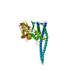
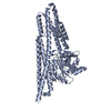
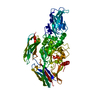
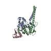
| 構造ビューア | 分子: MolmilJmol/JSmol |
|---|
- ダウンロードとリンク
 SASDF92


-モデル
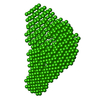
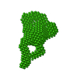
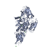
| モデル #2808 |   タイプ: dummy / ダミー原子の半径: 2.50 A コメント: Refined DAMMIN model from average volume (10 DAMMIF iterations) カイ2乗値: 0.955 / P-value: 0.891460  Omokage検索でこの集合体の類似形状データを探す (詳細) Omokage検索でこの集合体の類似形状データを探す (詳細) |
|---|
-試料
| 試料 | 名称: Urokinase plasminogen activator surface receptor, uPAR, T51C-V70C 試料濃度: 0.30-2.80 |
|---|---|
| バッファ | 名称: 20 mM PBS, 5 %(v/v) glycerol / pH: 7.4 |
| 要素 #66 | 名称: uPAR / タイプ: protein / 記述: Urokinase plasminogen activator surface receptor / 分子量: 36.98 / 分子数: 1 / 由来: Homo sapiens / 参照: UniProt: Q03405 配列: MGHPPLLPLL LLLHTCVPAS WGLRCMQCKT NGDCRVEECA LGQDLCRTTI VRLWEEGEEL ELVEKSCTHS EKTNRTLSYR TGLKITSLTE VVCGLDLCNQ GNSGRAVTYS RSRYLECISC GSSDMSCERG RHQSLQCRSP EEQCLDVVTH WIQEGEEGRP KDDRHLRGCG ...配列: MGHPPLLPLL LLLHTCVPAS WGLRCMQCKT NGDCRVEECA LGQDLCRTTI VRLWEEGEEL ELVEKSCTHS EKTNRTLSYR TGLKITSLTE VVCGLDLCNQ GNSGRAVTYS RSRYLECISC GSSDMSCERG RHQSLQCRSP EEQCLDVVTH WIQEGEEGRP KDDRHLRGCG YLPGCPGSNG FHNNDTFHFL KCCNTTKCNE GPILELENLP QNGRQCYSCK GNSTHGCSSE ETFLIDCRGP MNQCLVATGT HEPKNQSYMV RGCATASMCQ HAHLGDAFSM NHIDVSCCTK SGCNHPDLDV QYRSGAAPQP GPAHLSLTIT LLMTARLWGG TLLWT |
-実験情報
| ビーム | 設備名称: PETRA III EMBL P12 / 地域: Hamburg / 国: Germany / 線源: X-ray synchrotron / 波長: 0.124 Å / スペクトロメータ・検出器間距離: 3.1 mm | ||||||||||||||||||||||||||||||||||||||||||
|---|---|---|---|---|---|---|---|---|---|---|---|---|---|---|---|---|---|---|---|---|---|---|---|---|---|---|---|---|---|---|---|---|---|---|---|---|---|---|---|---|---|---|---|
| 検出器 | 名称: Pilatus 2M | ||||||||||||||||||||||||||||||||||||||||||
| スキャン |  測定日: 2017年12月1日 / 保管温度: 10 °C / セル温度: 10 °C / 照射時間: 0.05 sec. / フレーム数: 20 / 単位: 1/nm /
| ||||||||||||||||||||||||||||||||||||||||||
| 距離分布関数 P(R) | 
| ||||||||||||||||||||||||||||||||||||||||||
| 結果 |  コメント: Introduced disulfide in the first LU domain, DI, of uPAR (T51C-V70C).
|
