 Movie
Movie Controller
[English] 日本語
 Yorodumi
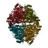
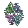
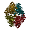
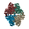
- SASDAX2: Pyruvate decarboxylase (PDC) from Z. mobilis (Pyruvate decarboxyl... -
+ Open data

- Basic information
| Entry | Database: SASBDB / ID: SASDAX2 |
|---|---|
 Sample Sample | Pyruvate decarboxylase (PDC) from Z. mobilis
|
| Function / homology |  Function and homology information Function and homology informationpyruvate decarboxylase / : / pyruvate decarboxylase activity / thiamine pyrophosphate binding / magnesium ion binding / cytosol Similarity search - Function |
| Biological species |  Zymomonas mobilis (bacteria) Zymomonas mobilis (bacteria) |
 Citation Citation | Journal: J Biol Chem / Year: 2000 Title: Crystal versus solution structures of thiamine diphosphate-dependent enzymes. Authors: D I Svergun / M V Petoukhov / M H Koch / S König /  Abstract: The quaternary structures of the thiamine diphosphate-dependent enzymes transketolase (EC 2.2.1.1; from Saccharomyces cerevisiae), pyruvate oxidase (EC 1.2.3.3; from Lactobacillus plantarum), and ...The quaternary structures of the thiamine diphosphate-dependent enzymes transketolase (EC 2.2.1.1; from Saccharomyces cerevisiae), pyruvate oxidase (EC 1.2.3.3; from Lactobacillus plantarum), and pyruvate decarboxylase (EC 4.1.1.1; from Zymomonas mobilis and brewers' yeast, the latter in the native and pyruvamide-activated forms) were examined by synchrotron x-ray solution scattering. The experimental scattering data were compared with the curves calculated from the crystallographic models of these multisubunit enzymes. For all enzymes noted above, except the very compact pyruvate decarboxylase from Z. mobilis, there were significant differences between the experimental and calculated profiles. The changes in relative positions of the subunits in solution were determined by rigid body refinement. For pyruvate oxidase and transketolase, which have tight intersubunit contacts in the crystal, relatively small modifications of the quaternary structure (root mean square displacements of 0.23 and 0.27 nm, respectively) sufficed to fit the experimental data. For the enzymes with looser contacts (the native and activated forms of yeast pyruvate decarboxylase), large modifications of the crystallographic models (root mean square displacements of 0.58 and 1.53 nm, respectively) were required. A clear correlation was observed between the magnitude of the distortions induced by the crystal environment and the interfacial area between subunits. |
 Contact author Contact author |
|
- Structure visualization
| Structure viewer | Molecule: MolmilJmol/JSmol |
|---|
- Downloads & links
-Data source
| SASBDB page |  SASDAX2 SASDAX2 |
|---|
-Related structure data
| Similar structure data |
|---|


-External links
| Related items in Molecule of the Month |
|---|


-Models
| Model #27 |  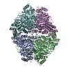 Type: atomic / Symmetry: P222 / Comment: This is Xtal model / Chi-square value: 0.665856 / P-value: 0.005737  Search similar-shape structures of this assembly by Omokage search (details) Search similar-shape structures of this assembly by Omokage search (details) |
|---|
-Sample
| Sample | Name: Pyruvate decarboxylase (PDC) from Z. mobilis / Sample MW: 244 kDa |
|---|---|
| Buffer | Name: 100 mM Sodium Citrate, 17% Glycerol, 22.5% PEG 1500 / pH: 6 / Comment: buffer for thiamin diphosphat dependent enzyme Composition: Glycerol 17.000 %, PEG 1500 22.500 %, Sodium Citrate 100.000 mM |
| Entity #20 | Name: ZmPDC / Type: protein / Description: Pyruvate decarboxylase / Formula weight: 60.93 / Num. of mol.: 4 / Source: Zymomonas mobilis / References: UniProt: P06672 Sequence: MSYTVGTYLA ERLVQIGLKH HFAVAGDYNL VLLDNLLLNK NMEQVYCCNE LNCGFSAEGY ARAKGAAAAV VTYSVGALSA FDAIGGAYAE NLPVILISGA PNNNDHAAGH VLHHALGKTD YHYQLEMAKN ITAAAEAIYT PEEAPAKIDH VIKTALREKK PVYLEIACNI ...Sequence: MSYTVGTYLA ERLVQIGLKH HFAVAGDYNL VLLDNLLLNK NMEQVYCCNE LNCGFSAEGY ARAKGAAAAV VTYSVGALSA FDAIGGAYAE NLPVILISGA PNNNDHAAGH VLHHALGKTD YHYQLEMAKN ITAAAEAIYT PEEAPAKIDH VIKTALREKK PVYLEIACNI ASMPCAAPGP ASALFNDEAS DEASLNAAVE ETLKFIANRD KVAVLVGSKL RAAGAEEAAV KFADALGGAV ATMAAAKSFF PEENPHYIGT SWGEVSYPGV EKTMKEADAV IALAPVFNDY STTGWTDIPD PKKLVLAEPR SVVVNGIRFP SVHLKDYLTR LAQKVSKKTG ALDFFKSLNA GELKKAAPAD PSAPLVNAEI ARQVEALLTP NTTVIAETGD SWFNAQRMKL PNGARVEYEM QWGHIGWSVP AAFGYAVGAP ERRNILMVGD GSFQLTAQEV AQMVRLKLPV IIFLINNYGY TIEVMIHDGP YNNIKNWDYA GLMEVFNGNG GYDSGAGKGL KAKTGGELAE AIKVALANTD GPTLIECFIG REDCTEELVK WGKRVAAANS RKPVNKLL |
-Experimental information
| Beam | Instrument name: DORIS III EMBL X33 / City: Hamburg / 国: Germany / Type of source: X-ray synchrotron | ||||||||||||
|---|---|---|---|---|---|---|---|---|---|---|---|---|---|
| Detector | Name: 1D Gas detector | ||||||||||||
| Scan | 
| ||||||||||||
| Distance distribution function P(R) | 
| ||||||||||||
| Result | 
|
