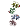
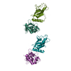
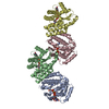
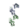
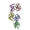
Journal: J Biol Chem / Year: 2013 Title: Conformational plasticity of the essential membrane-associated mannosyltransferase PimA from mycobacteria. Authors: David Giganti / Jorge Alegre-Cebollada / Saioa Urresti / David Albesa-Jové / Ane Rodrigo-Unzueta / Natalia Comino / Michael Kachala / Sonia López-Fernández / Dmitri I Svergun / Julio M ...Authors: David Giganti / Jorge Alegre-Cebollada / Saioa Urresti / David Albesa-Jové / Ane Rodrigo-Unzueta / Natalia Comino / Michael Kachala / Sonia López-Fernández / Dmitri I Svergun / Julio M Fernández / Marcelo E Guerin / Abstract: Phosphatidyl-myo-inositol mannosyltransferase A (PimA) is an essential glycosyltransferase (GT) that initiates the biosynthetic pathway of phosphatidyl-myo-inositol mannosides, lipomannan, and ...Phosphatidyl-myo-inositol mannosyltransferase A (PimA) is an essential glycosyltransferase (GT) that initiates the biosynthetic pathway of phosphatidyl-myo-inositol mannosides, lipomannan, and lipoarabinomannan, which are key glycolipids/lipoglycans of the mycobacterial cell envelope. PimA belongs to a large family of peripheral membrane-associated GTs for which the understanding of the molecular mechanism and conformational changes that govern substrate/membrane recognition and catalysis remains a major challenge. Here we used single molecule force spectroscopy techniques to study the mechanical and conformational properties of PimA. In our studies, we engineered a polyprotein containing PimA flanked by four copies of the well characterized I27 protein, which provides an unambiguous mechanical fingerprint. We found that PimA exhibits weak mechanical stability albeit displaying β-sheet topology expected to unfold at much higher forces. Notably, PimA unfolds following heterogeneous multiple step mechanical unfolding pathways at low force akin to molten globule states. Interestingly, the ab initio low resolution envelopes obtained from small angle x-ray scattering of the unliganded PimA and the PimA·GDP complexed forms clearly demonstrate that not only the "open" and "closed" conformations of the GT-B enzyme are largely present in solution, but in addition, PimA experiences remarkable flexibility that undoubtedly corresponds to the N-terminal "Rossmann fold" domain, which has been proved to participate in protein-membrane interactions. Based on these results and on our previous experimental data, we propose a model wherein the conformational transitions are important for the mannosyltransferase to interact with the donor and acceptor substrates/membrane.
Contact author
Michael Kachala (EMBL-Hamburg, European Molecular Biology Laboratory (EMBL) - Hamburg outstation, Notkestraße 85, Geb. 25A, 22607 Hamburg, Deutschland, Germany)
In the structure databanks used in Yorodumi, some data are registered as the other names, "COVID-19 virus" and "2019-nCoV". Here are the details of the virus and the list of structure data.
Jan 31, 2019. EMDB accession codes are about to change! (news from PDBe EMDB page)
EMDB accession codes are about to change! (news from PDBe EMDB page)
The allocation of 4 digits for EMDB accession codes will soon come to an end. Whilst these codes will remain in use, new EMDB accession codes will include an additional digit and will expand incrementally as the available range of codes is exhausted. The current 4-digit format prefixed with “EMD-” (i.e. EMD-XXXX) will advance to a 5-digit format (i.e. EMD-XXXXX), and so on. It is currently estimated that the 4-digit codes will be depleted around Spring 2019, at which point the 5-digit format will come into force.
The EM Navigator/Yorodumi systems omit the EMD- prefix.
Related info.:Q: What is EMD? / ID/Accession-code notation in Yorodumi/EM Navigator
Yorodumi is a browser for structure data from EMDB, PDB, SASBDB, etc.
This page is also the successor to EM Navigator detail page, and also detail information page/front-end page for Omokage search.
The word "yorodu" (or yorozu) is an old Japanese word meaning "ten thousand". "mi" (miru) is to see.
Related info.:EMDB / PDB / SASBDB / Comparison of 3 databanks / Yorodumi Search / Aug 31, 2016. New EM Navigator & Yorodumi / Yorodumi Papers / Jmol/JSmol / Function and homology information / Changes in new EM Navigator and Yorodumi
 Movie
Movie Controller

 Open data
 Basic information
 Sample
 Citation
 Contact author
 Structure visualization
 Molmil
 Downloads & links
 SASDAS4


 Search similar-shape structures of this assembly by Omokage search (details)
 / Type of source: X-ray synchrotron / Wavelength: 0.12 Å / Dist. spec. to detc.: 3.1 mm


