 Movie
Movie Controller
[English] 日本語
 Yorodumi
- SASDEF5: Phosphoketolase (L. lactis) with 1 mM thiaminpyrophosphate and 2%... -
+ Open data

- Basic information
| Entry | Database: SASBDB / ID: SASDEF5 |
|---|---|
 Sample Sample | Phosphoketolase (L. lactis) with 1 mM thiaminpyrophosphate and 2% v/v glycerol at pH 7.0
|
| Function / homology |  Function and homology information Function and homology informationLyases; Carbon-carbon lyases; Aldehyde-lyases / aldehyde-lyase activity / carbohydrate metabolic process Similarity search - Function |
| Biological species |  Lactococcus lactis subsp. lactis (strain IL1403) (lactic acid bacteria) Lactococcus lactis subsp. lactis (strain IL1403) (lactic acid bacteria) |
 Citation Citation | Journal: J Struct Biol / Year: 2019 Title: Crystal structure of a xylulose 5-phosphate phosphoketolase. Insights into the substrate specificity for xylulose 5-phosphate. Authors: A J Scheidig / D Horvath / S E Szedlacsek /    Abstract: Phosphoketolases (PK) are TPP-dependent enzymes which play essential roles in carbohydrate metabolism of numerous bacteria. Depending on the substrate specificity PKs can be subdivided into xylulose ...Phosphoketolases (PK) are TPP-dependent enzymes which play essential roles in carbohydrate metabolism of numerous bacteria. Depending on the substrate specificity PKs can be subdivided into xylulose 5-phosphate (X5P) specific PKs (XPKs) and PKs which accept both X5P and fructose 6-phosphate (F6P) (XFPKs). Despite their key metabolic importance, so far only the crystal structures of two XFPKs have been reported. There are no reported structures for any XPKs and for any complexes between PK and substrate. One of the major unknowns concerning PKs mechanism of action is related to the structural determinants of PKs substrate specificity for X5P or F6P. We report here the crystal structure of XPK from Lactococcus lactis (XPK-Ll) at 2.1 Å resolution. Using small angle X-ray scattering (SAXS) we proved that XPK-Ll is a dimer in solution. Towards better understanding of PKs substrate specificity, we performed flexible docking of TPP-X5P and TPP-F6P on crystal structures of XPK-Ll, two XFPKs and transketolase (TK). Calculated structure-based binding energies consistently support XPK-Ll preference for X5P. Analysis of structural models thus obtained show that substrates adopt moderately different conformation in PKs active sites following distinct networks of polar interactions. Based on the here reported structure of XPK-Ll we propose the most probable amino acid residues involved in the catalytic steps of reaction mechanism. Altogether our results suggest that PKs substrate preference for X5P or F6P is the outcome of a fine balance between specific binding network and dissimilar catalytic residues depending on the enzyme (XPK or XFPK) - substrate (X5P or F6P) couples. |
 Contact author Contact author |
|
- Structure visualization
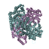
| Structure viewer | Molecule: MolmilJmol/JSmol |
|---|
- Downloads & links
-Data source
| SASBDB page |  SASDEF5 SASDEF5 |
|---|
-Related structure data


-External links
| Related items in Molecule of the Month |
|---|


-Models
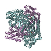
| Model #2447 |   Type: atomic / Symmetry: P2 Comment: PDB-ID 6GUA is a tetramer of dimers; the dimer is supposed to be the biological assembly in solution Chi-square value: 3.244  Search similar-shape structures of this assembly by Omokage search (details) Search similar-shape structures of this assembly by Omokage search (details) |
|---|---|
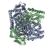
| Model #2448 |   Type: atomic / Symmetry: P1 / Chi-square value: 1.90 Search similar-shape structures of this assembly by Omokage search (details) |
-Sample
| Sample | Name: Phosphoketolase (L. lactis) with 1 mM thiaminpyrophosphate and 2% v/v glycerol at pH 7.0 Specimen concentration: 8 mg/ml |
|---|---|
| Buffer | Name: 20mM potassium phosphate 150mM NaCl 0.007 %(w/v) β-octyl glucoside 1mM DTT 1mM MgCl 1mM thiaminpyrophosphate 2 %(v/v) glycerol pH: 7 / Comment: SEC11 buffer |
| Entity #1316 | Type: protein / Description: Probable phosphoketolase / Formula weight: 95.727 / Num. of mol.: 2 / Source: Lactococcus lactis subsp. lactis (strain IL1403) / References: UniProt: Q9CFH4 Sequence: MSHHHHHHSM DIEFVDLEGS MTEYNSEAYL KKLDKWWRAA TYLGAGMIFL KENPLFSVTG TPIKAENLKA NPIGHWGTVS GQTFLYAHAN RLINKYDQKM FYMGGPGHGG QAMVVPSYLD GSYTEAYPEI TQDLEGMSRL FKRFSFPGGI GSHMTAQTPG SLHEGGELGY ...Sequence: MSHHHHHHSM DIEFVDLEGS MTEYNSEAYL KKLDKWWRAA TYLGAGMIFL KENPLFSVTG TPIKAENLKA NPIGHWGTVS GQTFLYAHAN RLINKYDQKM FYMGGPGHGG QAMVVPSYLD GSYTEAYPEI TQDLEGMSRL FKRFSFPGGI GSHMTAQTPG SLHEGGELGY VLSHATGAIL DQPEQIAFAV VGDGEAETGP LMTSWHSIKF INPKNDGAIL PILDLNGFKI SNPTLFARTS DVDIRKFFEG LGYSPRYIEN DDIHDYMAYH KLAAEVFDKA IEDIHQIQKD AREDNRYQNG EIPAWPIVIA RLPKGWGGPR YNDWSGPKFD GKGMPIEHSF RAHQVPLPLS SKNMGTLPEF VKWMTSYQPE TLFNADGSLK EELRDFAPKG EMRMASNPVT NGGVDSSNLV LPDWQEFANP ISENNRGKLL PDTNDNMDMN VLSKYFAEIV KLNPTRFRLF GPDETMSNRF WEMFKVTNRQ WMQVIKNPND EFISPEGRII DSQLSEHQAE GWLEGYTLTG RTGAFASYES FLRVVDSMLT QHFKWIRQAA DQKWRHDYPS LNVISTSTVF QQDHNGYTHQ DPGMLTHLAE KKSDFIRQYL PADGNTLLAV FDRAFQDRSK INHIVASKQP RQQWFTKEEA EKLATDGIAT IDWASTAKDG EAVDLVFASA GAEPTIETLA ALHLVNEVFP QAKFRYVNVV ELGRLQKKKG ALNQERELSD EEFEKYFGPS GTPVIFGFHG YEDLIESIFY QRGHDGLIVH GYREDGDITT TYDMRVYSEL DRFHQAIDAM QVLYVNRKVN QGLAKAFIDR MKRTLVKHFE VTRNEGVDIP DFTEWVWSDL KK |
-Experimental information
| Beam | Instrument name: PETRA III EMBL P12 / City: Hamburg / 国: Germany / Type of source: X-ray synchrotron / Wavelength: 0.124 Å / Dist. spec. to detc.: 3 mm | |||||||||||||||||||||||||||||||||
|---|---|---|---|---|---|---|---|---|---|---|---|---|---|---|---|---|---|---|---|---|---|---|---|---|---|---|---|---|---|---|---|---|---|---|
| Detector | Name: Pilatus 2M | |||||||||||||||||||||||||||||||||
| Scan |  Measurement date: Sep 7, 2017 / Storage temperature: 20 °C / Cell temperature: 20 °C / Exposure time: 0.045 sec. / Number of frames: 29 / Unit: 1/nm /
| |||||||||||||||||||||||||||||||||
| Distance distribution function P(R) | 
| |||||||||||||||||||||||||||||||||
| Result | 
|
