 Movie
Movie Controller
 Structure viewers
 About Yorodumi Papers


+Search query
-Structure paper
| Title | Crystal structure of a xylulose 5-phosphate phosphoketolase. Insights into the substrate specificity for xylulose 5-phosphate. |
|---|---|
| Journal, issue, pages | J Struct Biol, Vol. 207, Issue 1, Page 85-8102, Year 2019 |
| Publish date | Jul 1, 2019 |
 Authors Authors | A J Scheidig / D Horvath / S E Szedlacsek /    |
| PubMed Abstract | Phosphoketolases (PK) are TPP-dependent enzymes which play essential roles in carbohydrate metabolism of numerous bacteria. Depending on the substrate specificity PKs can be subdivided into xylulose ...Phosphoketolases (PK) are TPP-dependent enzymes which play essential roles in carbohydrate metabolism of numerous bacteria. Depending on the substrate specificity PKs can be subdivided into xylulose 5-phosphate (X5P) specific PKs (XPKs) and PKs which accept both X5P and fructose 6-phosphate (F6P) (XFPKs). Despite their key metabolic importance, so far only the crystal structures of two XFPKs have been reported. There are no reported structures for any XPKs and for any complexes between PK and substrate. One of the major unknowns concerning PKs mechanism of action is related to the structural determinants of PKs substrate specificity for X5P or F6P. We report here the crystal structure of XPK from Lactococcus lactis (XPK-Ll) at 2.1 Å resolution. Using small angle X-ray scattering (SAXS) we proved that XPK-Ll is a dimer in solution. Towards better understanding of PKs substrate specificity, we performed flexible docking of TPP-X5P and TPP-F6P on crystal structures of XPK-Ll, two XFPKs and transketolase (TK). Calculated structure-based binding energies consistently support XPK-Ll preference for X5P. Analysis of structural models thus obtained show that substrates adopt moderately different conformation in PKs active sites following distinct networks of polar interactions. Based on the here reported structure of XPK-Ll we propose the most probable amino acid residues involved in the catalytic steps of reaction mechanism. Altogether our results suggest that PKs substrate preference for X5P or F6P is the outcome of a fine balance between specific binding network and dissimilar catalytic residues depending on the enzyme (XPK or XFPK) - substrate (X5P or F6P) couples. |
 External links External links | J Struct Biol / PubMed:31059775 |
| Methods | SAS (X-ray synchrotron) / X-ray diffraction / small angle scattering |
| Resolution | 1.95 Å |
| Structure data | 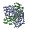 SASDEF5:  PDB-6gua: |
| Chemicals |  ChemComp-TPP:  ChemComp-MG:  ChemComp-PO4:  ChemComp-HOH: |
| Source |
|
 Keywords Keywords | LYASE / THIAMINE DIPHOSPHATE-DEPENDENT ENZYME / ALPHA-BETA FOLD / HOMODIMER |
 Lactococcus lactis subsp. lactis (strain il1403) (lactic acid bacteria)