 ムービー
ムービー コントローラー

+ データを開く

- 基本情報
基本情報
| 登録情報 | データベース: SASBDB / ID: SASDEL3 |
|---|---|
 試料 試料 | The neutrophil cytosol factor 2 (p67phox) subunit of phagocyte NADPH oxidase
|
| 機能・相同性 |  機能・相同性情報 機能・相同性情報superoxide-generating NADPH oxidase activator activity / phagolysosome / superoxide-generating NAD(P)H oxidase activity / Cross-presentation of particulate exogenous antigens (phagosomes) / NADPH oxidase complex / respiratory burst / ROS and RNS production in phagocytes / superoxide anion generation / superoxide metabolic process / Detoxification of Reactive Oxygen Species ...superoxide-generating NADPH oxidase activator activity / phagolysosome / superoxide-generating NAD(P)H oxidase activity / Cross-presentation of particulate exogenous antigens (phagosomes) / NADPH oxidase complex / respiratory burst / ROS and RNS production in phagocytes / superoxide anion generation / superoxide metabolic process / Detoxification of Reactive Oxygen Species / RHO GTPases Activate NADPH Oxidases / RAC2 GTPase cycle / RAC3 GTPase cycle / phagocytosis / cellular defense response / RAC1 GTPase cycle / acrosomal vesicle / small GTPase binding / VEGFA-VEGFR2 Pathway / electron transfer activity / innate immune response / membrane / plasma membrane / cytosol 類似検索 - 分子機能 |
| 生物種 |  Homo sapiens (ヒト) Homo sapiens (ヒト) |
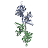
 引用 引用 | ジャーナル: J Biol Chem / 年: 2019 タイトル: Quantitative live-cell imaging and 3D modeling reveal critical functional features in the cytosolic complex of phagocyte NADPH oxidase. 著者: Cornelia S Ziegler / Leïla Bouchab / Marc Tramier / Dominique Durand / Franck Fieschi / Sophie Dupré-Crochet / Fabienne Mérola / Oliver Nüße / Marie Erard /  要旨: Phagocyte NADPH oxidase produces superoxide anions, a precursor of reactive oxygen species (ROS) critical for host responses to microbial infections. However, uncontrolled ROS production contributes ...Phagocyte NADPH oxidase produces superoxide anions, a precursor of reactive oxygen species (ROS) critical for host responses to microbial infections. However, uncontrolled ROS production contributes to inflammation, making NADPH oxidase a major drug target. It consists of two membranous (Nox2 and p22) and three cytosolic subunits (p40, p47, and p67) that undergo structural changes during enzyme activation. Unraveling the interactions between these subunits and the resulting conformation of the complex could shed light on NADPH oxidase regulation and help identify inhibition sites. However, the structures and the interactions of flexible proteins comprising several well-structured domains connected by intrinsically disordered protein segments are difficult to investigate by conventional techniques such as X-ray crystallography, NMR, or cryo-EM. Here, we developed an analytical strategy based on FRET-fluorescence lifetime imaging (FLIM) and fluorescence cross-correlation spectroscopy (FCCS) to structurally and quantitatively characterize NADPH oxidase in live cells. We characterized the inter- and intramolecular interactions of its cytosolic subunits by elucidating their conformation, stoichiometry, interacting fraction, and affinities in live cells. Our results revealed that the three subunits have a 1:1:1 stoichiometry and that nearly 100% of them are present in complexes in living cells. Furthermore, combining FRET data with small-angle X-ray scattering (SAXS) models and published crystal structures of isolated domains and subunits, we built a 3D model of the entire cytosolic complex. The model disclosed an elongated complex containing a flexible hinge separating two domains ideally positioned at one end of the complex and critical for oxidase activation and interactions with membrane components. |
 登録者 登録者 |
|
- 構造の表示
構造の表示
| 構造ビューア | 分子: MolmilJmol/JSmol |
|---|
- ダウンロードとリンク
 SASDEL3
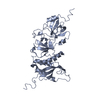


-モデル
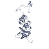
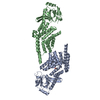
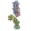
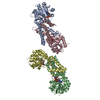
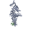
| モデル #2310 |   タイプ: atomic / カイ2乗値: 1.304 / P-value: 0.000001  Omokage検索でこの集合体の類似形状データを探す (詳細) Omokage検索でこの集合体の類似形状データを探す (詳細) |
|---|
-試料
| 試料 | 名称: The neutrophil cytosol factor 2 (p67phox) subunit of phagocyte NADPH oxidase 試料濃度: 0.30-3.80 |
|---|---|
| バッファ | 名称: 20 mM HEPES, 50 mM NaCl, 1 mM EDTA, 2 mM DTT, 5% glycerol pH: 8 |
| 要素 #1263 | 名称: p67phox / タイプ: protein / 記述: Neutrophil cytosol factor 2 / 分子量: 60.666 / 分子数: 1 / 由来: Homo sapiens / 参照: UniProt: P19878 配列: GPLGSPNSAR MSLVEAISLW NEGVLAADKK DWKGALDAFS AVQDPHSRIC FNIGCMYTIL KNMTEAEKAF TRSINRDKHL AVAYFQRGML YYQTEKYDLA IKDLKEALIQ LRGNQLIDYK ILGLQFKLFA CEVLYNIAFM YAKKEEWKKA EEQLALATSM KSEPRHSKID ...配列: GPLGSPNSAR MSLVEAISLW NEGVLAADKK DWKGALDAFS AVQDPHSRIC FNIGCMYTIL KNMTEAEKAF TRSINRDKHL AVAYFQRGML YYQTEKYDLA IKDLKEALIQ LRGNQLIDYK ILGLQFKLFA CEVLYNIAFM YAKKEEWKKA EEQLALATSM KSEPRHSKID KAMECVWKQK LYEPVVIPVG KLFRPNERQV AQLAKKDYLG KATVVASVVD QDSFSGFAPL QPQAAEPPPR PKTPEIFRAL EGEAHRVLFG FVPETKEELQ VMPGNIVFVL KKGNDNWATV MFNGQKGLVP CNYLEPVELR IHPQQQPQEE SSPQSDIPAP PSSKAPGRPQ LSPGQKQKEE PKEVKLSVPM PYTLKVHYKY TVVMKTQPGL PYSQVRDMVS KKLELRLEHT KLSYRPRDSN ELVPLSEDSM KDAWGQVKNY CLTLWCENTV GDQGFPDEPK ESEKADANNQ TTEPQLKKGS QVEALFSYEA TQPEDLEFQE GDIILVLSKV NEEWLEGESK GKVGIFPKVF VEDSATTDLE STRREV |
-実験情報
| ビーム | 設備名称: LURE D24 / 地域: Orsay / 国: France / 線源: X-ray synchrotron / 波長: 0.1488 Å / スペクトロメータ・検出器間距離: 1.378 mm | ||||||||||||||||||||||||||||||||||||||||||
|---|---|---|---|---|---|---|---|---|---|---|---|---|---|---|---|---|---|---|---|---|---|---|---|---|---|---|---|---|---|---|---|---|---|---|---|---|---|---|---|---|---|---|---|
| 検出器 | 名称: Custom / タイプ: Linear gas detector / Pixsize x: 605 mm | ||||||||||||||||||||||||||||||||||||||||||
| スキャン |  測定日: 2003年4月9日 / 保管温度: 4 °C / セル温度: 4 °C / 照射時間: 200 sec. / フレーム数: 8 / 単位: 1/A /
| ||||||||||||||||||||||||||||||||||||||||||
| 距離分布関数 P(R) | 
| ||||||||||||||||||||||||||||||||||||||||||
| 結果 | 
|
