 Movie
Movie Controller
 Structure viewers
 About Yorodumi Papers


+Search query
-Structure paper
| Title | Quantitative live-cell imaging and 3D modeling reveal critical functional features in the cytosolic complex of phagocyte NADPH oxidase. |
|---|---|
| Journal, issue, pages | J Biol Chem, Vol. 294, Issue 11, Page 3824-3836, Year 2019 |
| Publish date | Mar 15, 2019 |
 Authors Authors | Cornelia S Ziegler / Leïla Bouchab / Marc Tramier / Dominique Durand / Franck Fieschi / Sophie Dupré-Crochet / Fabienne Mérola / Oliver Nüße / Marie Erard /  |
| PubMed Abstract | Phagocyte NADPH oxidase produces superoxide anions, a precursor of reactive oxygen species (ROS) critical for host responses to microbial infections. However, uncontrolled ROS production contributes ...Phagocyte NADPH oxidase produces superoxide anions, a precursor of reactive oxygen species (ROS) critical for host responses to microbial infections. However, uncontrolled ROS production contributes to inflammation, making NADPH oxidase a major drug target. It consists of two membranous (Nox2 and p22) and three cytosolic subunits (p40, p47, and p67) that undergo structural changes during enzyme activation. Unraveling the interactions between these subunits and the resulting conformation of the complex could shed light on NADPH oxidase regulation and help identify inhibition sites. However, the structures and the interactions of flexible proteins comprising several well-structured domains connected by intrinsically disordered protein segments are difficult to investigate by conventional techniques such as X-ray crystallography, NMR, or cryo-EM. Here, we developed an analytical strategy based on FRET-fluorescence lifetime imaging (FLIM) and fluorescence cross-correlation spectroscopy (FCCS) to structurally and quantitatively characterize NADPH oxidase in live cells. We characterized the inter- and intramolecular interactions of its cytosolic subunits by elucidating their conformation, stoichiometry, interacting fraction, and affinities in live cells. Our results revealed that the three subunits have a 1:1:1 stoichiometry and that nearly 100% of them are present in complexes in living cells. Furthermore, combining FRET data with small-angle X-ray scattering (SAXS) models and published crystal structures of isolated domains and subunits, we built a 3D model of the entire cytosolic complex. The model disclosed an elongated complex containing a flexible hinge separating two domains ideally positioned at one end of the complex and critical for oxidase activation and interactions with membrane components. |
 External links External links | J Biol Chem / PubMed:30630949 / PubMed Central |
| Methods | SAS (X-ray in house) / SAS (X-ray synchrotron) |
| Structure data | 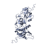 SASDEJ3: 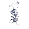 SASDEK3:  SASDEL3: |
| Source |
|
 Homo sapiens (human)