 Movie
Movie Controller
[English] 日本語
 Yorodumi
- SASDEH3: TubR protein of the pXO1-like plasmid pBc10987 from B. cereus (Bc... -
+ Open data

- Basic information
| Entry | Database: SASBDB / ID: SASDEH3 |
|---|---|
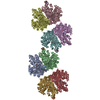
 Sample Sample | TubR protein of the pXO1-like plasmid pBc10987 from B. cereus (Bc-TubR) bound to S48 DNA (Bc-TubR : S48 DNA complex)
|
| Function / homology | Tubulin/FtsZ family, GTPase domain protein Function and homology information Function and homology information |
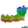
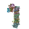
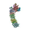
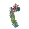
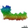
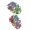
 Citation Citation | Journal: J Mol Biol / Year: 2018 Title: Cooperative DNA Binding of the Plasmid Partitioning Protein TubR from the Bacillus cereus pXO1 Plasmid. Authors: Ikuko Hayashi / Takashi Oda / Mamoru Sato / Sotaro Fuchigami /  Abstract: Tubulin/FtsZ-like GTPase TubZ is responsible for maintaining the stability of pXO1-like plasmids in virulent Bacilli. TubZ forms a filament in a GTP-dependent manner, and like other partitioning ...Tubulin/FtsZ-like GTPase TubZ is responsible for maintaining the stability of pXO1-like plasmids in virulent Bacilli. TubZ forms a filament in a GTP-dependent manner, and like other partitioning systems of low-copy-number plasmids, it requires the centromere-binding protein TubR that connects the plasmid to the TubZ filament. Systems regulating TubZ partitioning have been identified in Clostridium prophages as well as virulent Bacillus species, in which TubZ facilitates partitioning by binding and towing the segrosome: the nucleoprotein complex composed of TubR and the centromere. However, the molecular mechanisms of segrosome assembly and the transient on-off interactions between the segrosome and the TubZ filament remain poorly understood. Here, we determined the crystal structure of TubR from Bacillus cereus at 2.0-Å resolution and investigated the DNA-binding ability of TubR using hydroxyl radical footprinting and electrophoretic mobility shift assays. The TubR dimer possesses 2-fold symmetry and binds to a 15-bp palindromic consensus sequence in the tubRZ promoter region. Continuous TubR-binding sites overlap each other, which enables efficient binding of TubR in a cooperative manner. Interestingly, the segrosome adopts an extended DNA-protein filament structure and likely gains conformational flexibility by introducing non-consensus residues into the palindromes in an asymmetric manner. Together, our experimental results and structural model indicate that the unique centromere recognition mechanism of TubR allows transient complex formation between the segrosome and the dynamic polymer of TubZ. |
 Contact author Contact author |
|
- Structure visualization
| Structure viewer | Molecule: MolmilJmol/JSmol |
|---|
- Downloads & links
 SASDEH3
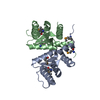
-Models
| Model #2304 |   Type: atomic  Search similar-shape structures of this assembly by Omokage search (details) Search similar-shape structures of this assembly by Omokage search (details) |
|---|---|
| Model #2305 | 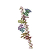 Type: atomic Search similar-shape structures of this assembly by Omokage search (details) |
| Model #2307 |  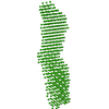 Type: dummy / Software: (DAMFILT 5.) / Radius of dummy atoms: 4.50 A / Chi-square value: 0.907 Search similar-shape structures of this assembly by Omokage search (details) |
-Sample
| Sample | Name: TubR protein of the pXO1-like plasmid pBc10987 from B. cereus (Bc-TubR) bound to S48 DNA (Bc-TubR : S48 DNA complex) Entity id: 1257 / 1258 / 1259 |
|---|---|
| Buffer | Name: 0.1 M NaCl, 10 mM Tris / pH: 8 |
| Entity #1257 | Type: DNA / Description: S48 DNA strand 1 / Formula weight: 21.324 / Num. of mol.: 1 Sequence: ATCATACTTC GGAAATATAT ACCGAAGTAT TTACGGCTTT TATAACGGTA TTAAATTCCG TATAATGAT |
| Entity #1258 | Type: DNA / Description: S48 DNA strand 2 / Formula weight: 21.324 / Num. of mol.: 1 Sequence: ATCATACTTC GGAAATATAT ACCGAAGTAT TTACGGCTTT TATAACGGTA TTAAATTCCG TATAATGAT |
| Entity #1259 | Name: Bc-TubR / Type: protein Description: TubR of the pXO1-like plasmid pBc10987 from B. cereus (Bc-TubR) Formula weight: 13.685 / Num. of mol.: 10 / References: UniProt: B7JTH0 Sequence: GSHMSNISMS SSEIIDVLCE NLNDGIWALR VLYAEGAMNK EKLWDYINQY HKDYQIENEK DYEGKKILPS RYALDIMTAR LEGAGLISFK AIGRVRIYDV TDLGNVLIKE LEKRVEKNN |
-Experimental information
| Beam | Instrument name: Photon Factory (PF), High Energy Accelerator Research Organization (KEK) BL-10C City: Tsukuba / 国: Japan / Type of source: X-ray synchrotron / Wavelength: 0.15 Å / Dist. spec. to detc.: 2.012 mm | ||||||||||||||||||||||||||||||
|---|---|---|---|---|---|---|---|---|---|---|---|---|---|---|---|---|---|---|---|---|---|---|---|---|---|---|---|---|---|---|---|
| Detector | Name: Pilatus3 2M / Pixsize x: 0.172 mm | ||||||||||||||||||||||||||||||
| Scan |  Measurement date: Nov 28, 2017 / Storage temperature: 8 °C / Cell temperature: 8 °C / Exposure time: 20 sec. / Number of frames: 1 / Unit: 1/A /
| ||||||||||||||||||||||||||||||
| Distance distribution function P(R) | 
| ||||||||||||||||||||||||||||||
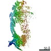
| Result |  Comments: The molecular weight (MW) of the Bc-TubR : S48 DNA complex was estimated from the macromolecular volume using the method of Fischer et al. (J. Appl. Crystallogr. 43, 101–109, 2010; SAXMow ...Comments: The molecular weight (MW) of the Bc-TubR : S48 DNA complex was estimated from the macromolecular volume using the method of Fischer et al. (J. Appl. Crystallogr. 43, 101–109, 2010; SAXMow program). However, the MW estimate is likely not an accurate value because the sample is a complex of protein and DNA. The theoretical SAXS curve of the molecular dynamics model was calculated as the mixture of the two models (one model contains four TubR dimers plus DNA, another model contains five TubR dimers plus DNA). For the mixed model, the fractions of each are 0.4 and 0.6, respectively. A low resolution dummy atom model was built by DAMMIF. Ten independently built models were generated, spatially aligned and then bead-occupancy and volume corrected using DAMAVER to generate an averaged spatial representation of the complex (damfilt). The the fit to the SAXS data from an individual dummy atom model from the cohort of models used to obtain the average representation is shown, specifically that with the smallest chi-square value. Sample concentration: UNKNOWN
|
