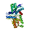
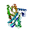
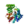
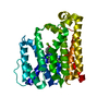
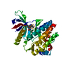
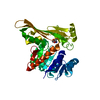
Journal: J Mol Biol / Year: 2018 Title: Cooperative DNA Binding of the Plasmid Partitioning Protein TubR from the Bacillus cereus pXO1 Plasmid. Authors: Ikuko Hayashi / Takashi Oda / Mamoru Sato / Sotaro Fuchigami / Abstract: Tubulin/FtsZ-like GTPase TubZ is responsible for maintaining the stability of pXO1-like plasmids in virulent Bacilli. TubZ forms a filament in a GTP-dependent manner, and like other partitioning ...Tubulin/FtsZ-like GTPase TubZ is responsible for maintaining the stability of pXO1-like plasmids in virulent Bacilli. TubZ forms a filament in a GTP-dependent manner, and like other partitioning systems of low-copy-number plasmids, it requires the centromere-binding protein TubR that connects the plasmid to the TubZ filament. Systems regulating TubZ partitioning have been identified in Clostridium prophages as well as virulent Bacillus species, in which TubZ facilitates partitioning by binding and towing the segrosome: the nucleoprotein complex composed of TubR and the centromere. However, the molecular mechanisms of segrosome assembly and the transient on-off interactions between the segrosome and the TubZ filament remain poorly understood. Here, we determined the crystal structure of TubR from Bacillus cereus at 2.0-Å resolution and investigated the DNA-binding ability of TubR using hydroxyl radical footprinting and electrophoretic mobility shift assays. The TubR dimer possesses 2-fold symmetry and binds to a 15-bp palindromic consensus sequence in the tubRZ promoter region. Continuous TubR-binding sites overlap each other, which enables efficient binding of TubR in a cooperative manner. Interestingly, the segrosome adopts an extended DNA-protein filament structure and likely gains conformational flexibility by introducing non-consensus residues into the palindromes in an asymmetric manner. Together, our experimental results and structural model indicate that the unique centromere recognition mechanism of TubR allows transient complex formation between the segrosome and the dynamic polymer of TubZ.
In the structure databanks used in Yorodumi, some data are registered as the other names, "COVID-19 virus" and "2019-nCoV". Here are the details of the virus and the list of structure data.
Jan 31, 2019. EMDB accession codes are about to change! (news from PDBe EMDB page)
EMDB accession codes are about to change! (news from PDBe EMDB page)
The allocation of 4 digits for EMDB accession codes will soon come to an end. Whilst these codes will remain in use, new EMDB accession codes will include an additional digit and will expand incrementally as the available range of codes is exhausted. The current 4-digit format prefixed with “EMD-” (i.e. EMD-XXXX) will advance to a 5-digit format (i.e. EMD-XXXXX), and so on. It is currently estimated that the 4-digit codes will be depleted around Spring 2019, at which point the 5-digit format will come into force.
The EM Navigator/Yorodumi systems omit the EMD- prefix.
Related info.:Q: What is EMD? / ID/Accession-code notation in Yorodumi/EM Navigator
Yorodumi is a browser for structure data from EMDB, PDB, SASBDB, etc.
This page is also the successor to EM Navigator detail page, and also detail information page/front-end page for Omokage search.
The word "yorodu" (or yorozu) is an old Japanese word meaning "ten thousand". "mi" (miru) is to see.
Related info.:EMDB / PDB / SASBDB / Comparison of 3 databanks / Yorodumi Search / Aug 31, 2016. New EM Navigator & Yorodumi / Yorodumi Papers / Jmol/JSmol / Function and homology information / Changes in new EM Navigator and Yorodumi
 Movie
Movie Controller


 Open data
 Basic information
 Components
 Keywords
 Function and homology information
 X-RAY DIFFRACTION /
  Authors
 Japan, 1items
  Citation
 Structure visualization
 Downloads & links
 Other downloads
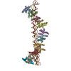


 PDBj
PDBj Assembly
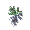
 Mass: 18.015 Da / Num. of mol.: 213 / Source method: isolated from a natural source / Formula: H2O
 Sample preparation
 Processing