Movie
Movie Controller


+ Open data

- Basic information
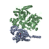
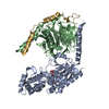
| Entry | Database: PDB / ID: 9yap | |||||||||||||||||||||
|---|---|---|---|---|---|---|---|---|---|---|---|---|---|---|---|---|---|---|---|---|---|---|
| Title | Gbg crosslinked to PLCb3 - second conformation | |||||||||||||||||||||
 Components Components |
| |||||||||||||||||||||
 Keywords Keywords | SIGNALING PROTEIN / G protein / heterotrimeric G protein / lipase / phospholipase | |||||||||||||||||||||
| Function / homology |  Function and homology information Function and homology informationphosphatidylinositol phospholipase C activity / regulation of systemic arterial blood pressure / phosphoinositide phospholipase C / Olfactory Signaling Pathway / Sensory perception of sweet, bitter, and umami (glutamate) taste / Fatty Acids bound to GPR40 (FFAR1) regulate insulin secretion / Synthesis, secretion, and inactivation of Glucagon-like Peptide-1 (GLP-1) / Acetylcholine regulates insulin secretion / phosphatidylinositol metabolic process / phospholipase C-activating serotonin receptor signaling pathway ...phosphatidylinositol phospholipase C activity / regulation of systemic arterial blood pressure / phosphoinositide phospholipase C / Olfactory Signaling Pathway / Sensory perception of sweet, bitter, and umami (glutamate) taste / Fatty Acids bound to GPR40 (FFAR1) regulate insulin secretion / Synthesis, secretion, and inactivation of Glucagon-like Peptide-1 (GLP-1) / Acetylcholine regulates insulin secretion / phosphatidylinositol metabolic process / phospholipase C-activating serotonin receptor signaling pathway / phosphatidylinositol-4,5-bisphosphate phospholipase C activity / PLC beta mediated events / C-type glycerophospholipase activity / Activation of the phototransduction cascade / Activation of G protein gated Potassium channels / G-protein activation / G beta:gamma signalling through PI3Kgamma / Prostacyclin signalling through prostacyclin receptor / G beta:gamma signalling through PLC beta / ADP signalling through P2Y purinoceptor 1 / Thromboxane signalling through TP receptor / Presynaptic function of Kainate receptors / G beta:gamma signalling through CDC42 / Inhibition of voltage gated Ca2+ channels via Gbeta/gamma subunits / G alpha (12/13) signalling events / Glucagon-type ligand receptors / G beta:gamma signalling through BTK / ADP signalling through P2Y purinoceptor 12 / Adrenaline,noradrenaline inhibits insulin secretion / Cooperation of PDCL (PhLP1) and TRiC/CCT in G-protein beta folding / Ca2+ pathway / G alpha (z) signalling events / Thrombin signalling through proteinase activated receptors (PARs) / Extra-nuclear estrogen signaling / G alpha (s) signalling events / G alpha (q) signalling events / Glucagon-like Peptide-1 (GLP1) regulates insulin secretion / G alpha (i) signalling events / High laminar flow shear stress activates signaling by PIEZO1 and PECAM1:CDH5:KDR in endothelial cells / Vasopressin regulates renal water homeostasis via Aquaporins / Synthesis of IP3 and IP4 in the cytosol / phosphatidylinositol-mediated signaling / postsynaptic cytosol / lipid catabolic process / release of sequestered calcium ion into cytosol / molecular function activator activity / G beta:gamma signalling through PLC beta / Presynaptic function of Kainate receptors / photoreceptor disc membrane / cellular response to catecholamine stimulus / adenylate cyclase-activating dopamine receptor signaling pathway / cellular response to prostaglandin E stimulus / heterotrimeric G-protein complex / G-protein beta-subunit binding / sensory perception of taste / signaling receptor complex adaptor activity / retina development in camera-type eye / GTPase binding / Ca2+ pathway / molecular adaptor activity / G alpha (q) signalling events / phospholipase C-activating G protein-coupled receptor signaling pathway / calmodulin binding / cell population proliferation / cadherin binding / G protein-coupled receptor signaling pathway / GTPase activity / calcium ion binding / synapse / protein-containing complex binding / protein-containing complex / membrane / nucleus / plasma membrane / cytosol / cytoplasm Similarity search - Function | |||||||||||||||||||||
| Biological species |  Homo sapiens (human) Homo sapiens (human) | |||||||||||||||||||||
| Method | ELECTRON MICROSCOPY / single particle reconstruction / cryo EM / Resolution: 4.5 Å | |||||||||||||||||||||
 Authors Authors | Fisher, I.J. / Lyon, A.M. | |||||||||||||||||||||
| Funding support |  United States, 3items United States, 3items
| |||||||||||||||||||||
 Citation Citation | Journal: bioRxiv / Year: 2026 Title: Gβγ engages PLCβ3 at multiple sites to reorient and facilitate its activation. Authors: Isaac J Fisher / Kanishka Senarath / Kennedy Outlaw / Kaushik Muralidharan / Elisabeth E Garland-Kuntz / Michelle Van Camp / Tommy Komay / Asuka Inoue / Eva Kostenis / Nevin A Lambert / Angeline M Lyon /   Abstract: Phospholipase C β (PLCβ) enzymes are activated by heterotrimeric G protein subunits, increasing hydrolysis of phosphatidylinositol-4,5-bisphosphate (PIP2) at the plasma membrane. All four human ...Phospholipase C β (PLCβ) enzymes are activated by heterotrimeric G protein subunits, increasing hydrolysis of phosphatidylinositol-4,5-bisphosphate (PIP2) at the plasma membrane. All four human PLCβ isoforms (PLCβ1-4) are activated by Gα, while PLCβ1-3 are activated to varying extents by Gβγ. The binding sites for Gα on PLCβ are well-established and much has been learned about its mechanism of activation, but comparatively little is known about Gβγ-dependent activation. In this work, we used cryo-electron microscopy (cryo-EM) single particle analysis (SPA), functional assays, and bioluminescence resonance energy transfer (BRET) to investigate how Gβγ interacts with PLCβ3 in concert with activated Gα to regulate phospholipase activity. Gβγ heterodimers bind multiple surfaces of PLCβ3 to promote activation but alone do not recruit the enzyme to the plasma membrane. Instead, Gβγ facilitates activation by Gα, most likely by reorienting the phospholipase catalytic site at the membrane to maximize PIP2 hydrolysis and downstream Ca release. Cell-based functional assays demonstrate that Gβγ is required for maximal PLCβ3 activation even when G heterotrimers are the sole source of Gβγ. Together, these findings demonstrate that Gβγ acts as a critical positive allosteric modulator that regularly acts in concert with Gα to activate PLCβ3 at the plasma membrane. | |||||||||||||||||||||
| History |
|
- Structure visualization
| Structure viewer | Molecule: MolmilJmol/JSmol |
|---|
- Downloads & links
-Download
| PDBx/mmCIF format | 9yap.cif.gz | 332.8 KB | Display | PDBx/mmCIF format |
|---|---|---|---|---|
| PDB format | pdb9yap.ent.gz | 268.5 KB | Display | PDB format |
| PDBx/mmJSON format | 9yap.json.gz | Tree view | PDBx/mmJSON format | |
| Others |  Other downloads Other downloads |
-Validation report
| Arichive directory | https://data.pdbj.org/pub/pdb/validation_reports/ya/9yapftp://data.pdbj.org/pub/pdb/validation_reports/ya/9yap | HTTPS FTP |
|---|
-Related structure data
| Related structure data |  72733MC  9y7hC  9yaoC C: citing same article ( M: map data used to model this data |
|---|---|
| Similar structure data |


-Links
 PDBj
PDBj


- Assembly
| Deposited unit | 
|
|---|---|
| 1 |
|
-Components
| #1: Protein | Mass: 99653.891 Da / Num. of mol.: 1 / Mutation: E60C, S193C, S221C, S358C, S824C, S834C Source method: isolated from a genetically manipulated source Source: (gene. exp.) Homo sapiens (human) / Gene: PLCB3 / Production host:   Spodoptera frugiperda (fall armyworm) Spodoptera frugiperda (fall armyworm)References: UniProt: Q01970, phosphoinositide phospholipase C |
|---|---|
| #2: Protein | Mass: 37285.734 Da / Num. of mol.: 1 Source method: isolated from a genetically manipulated source Source: (gene. exp.) Trichoplusia ni (cabbage looper) / References: UniProt: P62871 |
| #3: Protein | Mass: 7861.143 Da / Num. of mol.: 1 / Mutation: C68S Source method: isolated from a genetically manipulated source Source: (gene. exp.) Trichoplusia ni (cabbage looper) / References: UniProt: P63212 |
| Has protein modification | N |
-Experimental details
-Experiment
| Experiment | Method: ELECTRON MICROSCOPY |
|---|---|
| EM experiment | Aggregation state: PARTICLE / 3D reconstruction method: single particle reconstruction |
- Sample preparation
| Component | Name: BMPEG-crosslinked complex of Gb1g2 and PLCb3 / Type: COMPLEX / Entity ID: all / Source: MULTIPLE SOURCES |
|---|---|
| Molecular weight | Value: 0.156 MDa / Experimental value: NO |
| Source (natural) | Organism: Homo sapiens (human) |
| Source (recombinant) | Organism: Spodoptera frugiperda (fall armyworm) |
| Buffer solution | pH: 7.4 Details: 20 mM HEPES pH 7.4, 100 mM NaCl, 0.1 mM EDTA and 0.1 mM EGTA |
| Specimen | Conc.: 1 mg/ml / Embedding applied: NO / Shadowing applied: NO / Staining applied: NO / Vitrification applied: YES |
| Vitrification | Instrument: FEI VITROBOT MARK IV / Cryogen name: ETHANE |
- Electron microscopy imaging
| Experimental equipment |  Model: Titan Krios / Image courtesy: FEI Company |
|---|---|
| Microscopy | Model: TFS KRIOS |
| Electron gun | Electron source:  FIELD EMISSION GUN / Accelerating voltage: 300 kV / Illumination mode: FLOOD BEAM FIELD EMISSION GUN / Accelerating voltage: 300 kV / Illumination mode: FLOOD BEAM |
| Electron lens | Mode: BRIGHT FIELD / Nominal defocus max: 3000 nm / Nominal defocus min: 1000 nm |
| Image recording | Electron dose: 53.69 e/Å2 / Film or detector model: GATAN K3 BIOQUANTUM (6k x 4k) |
- Processing
| EM software |
| ||||||||||||||||||||||||||||||||||||||||
|---|---|---|---|---|---|---|---|---|---|---|---|---|---|---|---|---|---|---|---|---|---|---|---|---|---|---|---|---|---|---|---|---|---|---|---|---|---|---|---|---|---|
| CTF correction | Type: PHASE FLIPPING AND AMPLITUDE CORRECTION | ||||||||||||||||||||||||||||||||||||||||
| 3D reconstruction | Resolution: 4.5 Å / Resolution method: FSC 0.143 CUT-OFF / Num. of particles: 142175 / Symmetry type: POINT | ||||||||||||||||||||||||||||||||||||||||
| Atomic model building | Protocol: FLEXIBLE FIT Details: Crystal structures of Gbetagamma and PLCbeta3 (PDB IDs 1GP2 and 4GNK) were rigid-body fit into the cryo-EM map using Chimera. The model was refined using molecular dynamic flexible fitting ...Details: Crystal structures of Gbetagamma and PLCbeta3 (PDB IDs 1GP2 and 4GNK) were rigid-body fit into the cryo-EM map using Chimera. The model was refined using molecular dynamic flexible fitting (MDFF). MDFF configuration files were generated using VMD. During MDFF simulation, Gbetagamma was set as rigid with domain restraints. The MDFF simulation was conducted with a grid scaling value of 0.5 for 100 ps, followed by 3,000 steps of energy minimization until convergence of the protein RMSD. The MDFF generated model was inspected and manually adjusted in Coot, guided through the use of deep-learning-based amino-acid-wise model quality (DAQ) scoring, and refined in PHENIX. | ||||||||||||||||||||||||||||||||||||||||
| Atomic model building |
| ||||||||||||||||||||||||||||||||||||||||
| Refinement | Highest resolution: 4.5 Å Stereochemistry target values: REAL-SPACE (WEIGHTED MAP SUM AT ATOM CENTERS) | ||||||||||||||||||||||||||||||||||||||||
| Refine LS restraints |
|