 Movie
Movie Controller


+ Open data

- Basic information
| Entry | Database: PDB / ID: 9y0h | |||||||||||||||||||||
|---|---|---|---|---|---|---|---|---|---|---|---|---|---|---|---|---|---|---|---|---|---|---|
| Title | Insulin Degrading Enzyme Time-resolved O/O state | |||||||||||||||||||||
 Components Components | Cysteine-free Insulin-degrading enzyme | |||||||||||||||||||||
 Keywords Keywords | HYDROLASE / M16A zinc metalloprotease | |||||||||||||||||||||
| Function / homology |  Function and homology information Function and homology informationinsulysin / beta-endorphin binding / ubiquitin recycling / insulin catabolic process / insulin metabolic process / amyloid-beta clearance by cellular catabolic process / hormone catabolic process / bradykinin catabolic process / cytosolic proteasome complex / positive regulation of protein binding ...insulysin / beta-endorphin binding / ubiquitin recycling / insulin catabolic process / insulin metabolic process / amyloid-beta clearance by cellular catabolic process / hormone catabolic process / bradykinin catabolic process / cytosolic proteasome complex / positive regulation of protein binding / insulin binding / regulation of aerobic respiration / peptide catabolic process / peroxisomal matrix / amyloid-beta clearance / amyloid-beta metabolic process / Insulin receptor recycling / negative regulation of proteolysis / peptide binding / protein catabolic process / : / Peroxisomal protein import / antigen processing and presentation of endogenous peptide antigen via MHC class I / metalloendopeptidase activity / insulin receptor signaling pathway / positive regulation of protein catabolic process / peroxisome / amyloid-beta binding / virus receptor activity / endopeptidase activity / basolateral plasma membrane / Ub-specific processing proteases / external side of plasma membrane / protein-containing complex binding / cell surface / protein homodimerization activity / ATP hydrolysis activity / mitochondrion / proteolysis / : / extracellular exosome / zinc ion binding / ATP binding / identical protein binding / nucleus / cytosol / cytoplasm Similarity search - Function | |||||||||||||||||||||
| Biological species |  Homo sapiens (human) Homo sapiens (human) | |||||||||||||||||||||
| Method | ELECTRON MICROSCOPY / single particle reconstruction / cryo EM / Resolution: 5.1 Å | |||||||||||||||||||||
 Authors Authors | Mancl, J.M. / Tang, W.J. | |||||||||||||||||||||
| Funding support |  United States, 1items United States, 1items
| |||||||||||||||||||||
 Citation Citation | Journal: Elife / Year: 2026 Title: Characterization and modulation of human insulin degrading enzyme conformational dynamics to control enzyme activity. Authors: Jordan M Mancl / Wenguang G Liang / Nicholas L Bayhi / Hui Wei / William C Budell / Joshua H Mendez / Tobin R Sosnick / Bridget Carragher / Clinton S Potter / Wei-Jen Tang / Abstract: Insulin degrading enzyme (IDE) is a dimeric M16A zinc metalloprotease that degrades amyloidogenic peptides diverse in shape and sequence, including insulin and amyloid-β, to prevent toxic amyloid ...Insulin degrading enzyme (IDE) is a dimeric M16A zinc metalloprotease that degrades amyloidogenic peptides diverse in shape and sequence, including insulin and amyloid-β, to prevent toxic amyloid fibril formation. IDE has a hollow catalytic chamber formed by two ~55 kDa N- and C- domains (IDE-N and IDE-C, respectively), in which peptides bind, unfold, and are repositioned for proteolysis. IDE is known to transition between a closed state, poised for catalysis, and an open state, able to release cleavage products and bind a new substrate. Here, we present six cryo-EM structures of the IDE dimer at 3.0-5.1 Å resolution, obtained in the presence of a sub-saturating concentration of insulin. Combining cryo-EM heterogeneity analysis with all-atom molecular dynamics (MD) simulations, we identified the structural basis and key residues for IDE conformational dynamics that were not previously revealed by IDE static structures. Notably, R668 serves as a molecular latch mediating the open-close transition and facilitates key protein motions through charge-swapping interactions at the IDE-N/C interface. Our small-angle X-ray scattering analysis and enzymatic assays of an R668A mutant indicate a profound alteration of conformational dynamics and catalytic activity. By integrating coarse-grained MD simulations, our analysis reveals that IDE unfolds its substrates through the coordinated motion between IDE-N and IDE-C, as well as β-sheet formation between IDE and insulin. Additionally, our time-resolved cryo-EM analysis uncovers IDE allostery within the IDE dimer. Collectively, our findings demonstrate the strength of combining experimental and computational approaches to probe protein dynamics and pave the way for developing substrate-specific modulators of IDE activity. #1: Journal: eLife / Year: 2025Title: Characterization and modulation of human insulin degrading enzyme conformational dynamics to control enzyme activity Authors: Mancl, J.M. / Liang, W.G. / Wei, H. / Carragher, B. / Potter, C.S. / Tang, W.J. | |||||||||||||||||||||
| History |
|
- Structure visualization
| Structure viewer | Molecule: MolmilJmol/JSmol |
|---|
- Downloads & links
-Download
| PDBx/mmCIF format | 9y0h.cif.gz | 344.8 KB | Display | PDBx/mmCIF format |
|---|---|---|---|---|
| PDB format | pdb9y0h.ent.gz | 279.3 KB | Display | PDB format |
| PDBx/mmJSON format | 9y0h.json.gz | Tree view | PDBx/mmJSON format | |
| Others |  Other downloads Other downloads |
-Validation report
| Arichive directory | https://data.pdbj.org/pub/pdb/validation_reports/y0/9y0hftp://data.pdbj.org/pub/pdb/validation_reports/y0/9y0h | HTTPS FTP |
|---|
-Related structure data
| Related structure data |  72393MC M: map data used to model this data C: citing same article ( |
|---|---|
| Similar structure data |


-Links
 PDBj
PDBj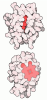
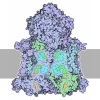
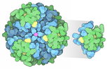
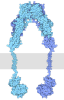

- Assembly
| Deposited unit | 
|
|---|---|
| 1 |
|
-Components
| #1: Protein | Mass: 117068.508 Da / Num. of mol.: 2 Source method: isolated from a genetically manipulated source Source: (gene. exp.) Homo sapiens (human) / Gene: IDE / Production host:   Escherichia coli (E. coli) / References: UniProt: P14735, insulysin Escherichia coli (E. coli) / References: UniProt: P14735, insulysinHas protein modification | N | |
|---|
-Experimental details
-Experiment
| Experiment | Method: ELECTRON MICROSCOPY |
|---|---|
| EM experiment | Aggregation state: PARTICLE / 3D reconstruction method: single particle reconstruction |
- Sample preparation
| Component | Name: Cysteine-free Insulin-degrading enzyme / Type: COMPLEX / Entity ID: all / Source: RECOMBINANT |
|---|---|
| Source (natural) | Organism: Homo sapiens (human) |
| Source (recombinant) | Organism: Escherichia coli (E. coli) |
| Buffer solution | pH: 7.5 |
| Specimen | Embedding applied: NO / Shadowing applied: NO / Staining applied: NO / Vitrification applied: YES |
| Vitrification | Instrument: SPOTITON / Cryogen name: ETHANE |
- Electron microscopy imaging
| Experimental equipment |  Model: Titan Krios / Image courtesy: FEI Company |
|---|---|
| Microscopy | Model: TFS KRIOS |
| Electron gun | Electron source:  FIELD EMISSION GUN / Accelerating voltage: 300 kV / Illumination mode: FLOOD BEAM FIELD EMISSION GUN / Accelerating voltage: 300 kV / Illumination mode: FLOOD BEAM |
| Electron lens | Mode: BRIGHT FIELD / Nominal defocus max: 2500 nm / Nominal defocus min: 700 nm |
| Image recording | Electron dose: 65 e/Å2 / Film or detector model: GATAN K3 (6k x 4k) |
| EM imaging optics | Energyfilter slit width: 20 eV |
- Processing
| EM software |
| ||||||||||||||||||||||||
|---|---|---|---|---|---|---|---|---|---|---|---|---|---|---|---|---|---|---|---|---|---|---|---|---|---|
| CTF correction | Type: PHASE FLIPPING AND AMPLITUDE CORRECTION | ||||||||||||||||||||||||
| 3D reconstruction | Resolution: 5.1 Å / Resolution method: FSC 0.143 CUT-OFF / Num. of particles: 376750 / Symmetry type: POINT | ||||||||||||||||||||||||
| Refinement | Highest resolution: 5.1 Å Stereochemistry target values: REAL-SPACE (WEIGHTED MAP SUM AT ATOM CENTERS) | ||||||||||||||||||||||||
| Refine LS restraints |
|
