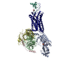
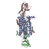
Movie
Movie Controller

[English] 日本語
 Yorodumi
- PDB-9xjp: Structure of the CX3CL1.44-US28-GqiN18-scFv16 in the E-state, che... -
+ Open data

- Basic information
| Entry | Database: PDB / ID: 9xjp | |||||||||
|---|---|---|---|---|---|---|---|---|---|---|
| Title | Structure of the CX3CL1.44-US28-GqiN18-scFv16 in the E-state, chemokine domain fully modeled | |||||||||
 Components Components |
| |||||||||
 Keywords Keywords | MEMBRANE PROTEIN / GPCR / intermediate state / GDP / engineered chemokine / HCMV / viral protein / viral-host interaction | |||||||||
| Function / homology |  Function and homology information Function and homology informationCXCR1 chemokine receptor binding / positive regulation of calcium-independent cell-cell adhesion / leukocyte adhesive activation / CX3C chemokine receptor binding / negative regulation of glutamate receptor signaling pathway / negative regulation of interleukin-1 alpha production / negative regulation of neuron migration / positive regulation of microglial cell migration / regulation of lipopolysaccharide-mediated signaling pathway / negative regulation of microglial cell activation ...CXCR1 chemokine receptor binding / positive regulation of calcium-independent cell-cell adhesion / leukocyte adhesive activation / CX3C chemokine receptor binding / negative regulation of glutamate receptor signaling pathway / negative regulation of interleukin-1 alpha production / negative regulation of neuron migration / positive regulation of microglial cell migration / regulation of lipopolysaccharide-mediated signaling pathway / negative regulation of microglial cell activation / microglial cell proliferation / negative regulation of hippocampal neuron apoptotic process / positive regulation of gonadotropin secretion / autocrine signaling / phospholipase C-activating tachykinin receptor signaling pathway / Fatty Acids bound to GPR40 (FFAR1) regulate insulin secretion / Acetylcholine regulates insulin secretion / CCR chemokine receptor binding / leukocyte migration involved in inflammatory response / phospholipase C-activating G protein-coupled glutamate receptor signaling pathway / positive regulation of actin filament bundle assembly / regulation of platelet activation / synapse pruning / PLC beta mediated events / phospholipase C-activating serotonin receptor signaling pathway / entrainment of circadian clock / sensory perception of itch / positive regulation of membrane depolarization / integrin activation / phototransduction, visible light / C-C chemokine binding / eosinophil chemotaxis / C-C chemokine receptor activity / positive regulation of cell-matrix adhesion / neuron cellular homeostasis / positive regulation of neuroblast proliferation / chemokine activity / leukocyte chemotaxis / Chemokine receptors bind chemokines / regulation of canonical Wnt signaling pathway / negative regulation of interleukin-1 beta production / symbiont-mediated transformation of host cell / neuron remodeling / negative regulation of interleukin-6 production / positive chemotaxis / chemoattractant activity / cell projection / glutamate receptor signaling pathway / negative regulation of apoptotic signaling pathway / negative regulation of cell-substrate adhesion / negative regulation of tumor necrosis factor production / regulation of neurogenesis / postsynaptic cytosol / photoreceptor outer segment / response to ischemia / hormone-mediated signaling pathway / cellular response to acidic pH / symbiont-mediated perturbation of host defense response / regulation of eating behavior / adenylate cyclase inhibitor activity / T cell migration / positive regulation of protein localization to cell cortex / positive regulation of relaxation of smooth muscle / Adenylate cyclase inhibitory pathway / positive regulation of smooth muscle cell proliferation / D2 dopamine receptor binding / adenylate cyclase-inhibiting dopamine receptor signaling pathway / GTPase activator activity / negative regulation of cell migration / adenylate cyclase-inhibiting serotonin receptor signaling pathway / G protein-coupled serotonin receptor binding / Turbulent (oscillatory, disturbed) flow shear stress activates signaling by PIEZO1 and integrins in endothelial cells / regulation of G protein-coupled receptor signaling pathway / positive regulation of release of sequestered calcium ion into cytosol / cellular response to forskolin / regulation of mitotic spindle organization / mast cell degranulation / chemokine-mediated signaling pathway / calcium-mediated signaling / positive regulation of neuron projection development / cell chemotaxis / establishment of mitotic spindle orientation / defense response / cell-cell adhesion / neuropeptide signaling pathway / Regulation of insulin secretion / response to prostaglandin E / microglial cell activation / regulation of synaptic plasticity / positive regulation of cholesterol biosynthetic process / G protein-coupled receptor binding / integrin binding / blood coagulation / chemotaxis / response to peptide hormone / cytokine-mediated signaling pathway / G-protein beta/gamma-subunit complex binding / adenylate cyclase-inhibiting G protein-coupled receptor signaling pathway / adenylate cyclase-modulating G protein-coupled receptor signaling pathway / positive regulation of inflammatory response Similarity search - Function | |||||||||
| Biological species |  Homo sapiens (human) Homo sapiens (human)  Mus musculus (house mouse) Mus musculus (house mouse)  Human betaherpesvirus 5 Human betaherpesvirus 5 | |||||||||
| Method | ELECTRON MICROSCOPY / single particle reconstruction / cryo EM / Resolution: 2.74 Å | |||||||||
 Authors Authors | Jude, K.M. / Garcia, K.C. / Tsutsumi, N. | |||||||||
| Funding support |  Japan, 1items Japan, 1items
| |||||||||
 Citation Citation | Journal: To Be Published Title: Structural basis of a stepwise Gq activation by a chemokine receptor US28 Authors: Jude, K.M. / Garcia, K.C. / Tsutsumi, N. | |||||||||
| History |
|
- Structure visualization
| Structure viewer | Molecule: MolmilJmol/JSmol |
|---|
- Downloads & links
-Download
| PDBx/mmCIF format | 9xjp.cif.gz | 348.5 KB | Display | PDBx/mmCIF format |
|---|---|---|---|---|
| PDB format | pdb9xjp.ent.gz | 219.9 KB | Display | PDB format |
| PDBx/mmJSON format | 9xjp.json.gz | Tree view | PDBx/mmJSON format | |
| Others |  Other downloads Other downloads |
-Validation report
| Arichive directory | https://data.pdbj.org/pub/pdb/validation_reports/xj/9xjpftp://data.pdbj.org/pub/pdb/validation_reports/xj/9xjp | HTTPS FTP |
|---|
-Related structure data
| Related structure data |  66942MC  9xh2C  9xixC  9xiyC  9xj5C  9xj7C C: citing same article ( M: map data used to model this data |
|---|---|
| Similar structure data |


-Links
 PDBj
PDBj













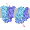




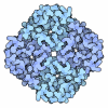





- Assembly
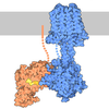
| Deposited unit | 
|
|---|---|
| 1 |
|
-Components
-Guanine nucleotide-binding protein ... , 3 types, 3 molecules ABC
| #1: Protein | Mass: 41249.934 Da / Num. of mol.: 1 Source method: isolated from a genetically manipulated source Details: residues 1-25 MTLESIMACCLSEEAKEARRINDEI are replaced with GCTLSAEDKAAVERSKMI from G(i),residues 1-25 MTLESIMACCLSEEAKEARRINDEI are replaced with GCTLSAEDKAAVERSKMI from G(i) Source: (gene. exp.) Homo sapiens (human) / Gene: GNAI1, GNAQ, GAQ / Production host:  Trichoplusia ni (cabbage looper) Trichoplusia ni (cabbage looper)References: UniProt: P63096, UniProt: P50148, Hydrolases; Acting on acid anhydrides; Acting on GTP to facilitate cellular and subcellular movement |
|---|---|
| #2: Protein | Mass: 37728.152 Da / Num. of mol.: 1 Source method: isolated from a genetically manipulated source Source: (gene. exp.) Homo sapiens (human) / Gene: GNB1 / Production host: Trichoplusia ni (cabbage looper) / References: UniProt: P62873 |
| #3: Protein | Mass: 7432.554 Da / Num. of mol.: 1 Source method: isolated from a genetically manipulated source Source: (gene. exp.) Homo sapiens (human) / Gene: GNG2 / Production host: Trichoplusia ni (cabbage looper) / References: UniProt: P59768 |
-Protein , 2 types, 2 molecules RL
| #5: Protein | Mass: 42041.098 Da / Num. of mol.: 1 Source method: isolated from a genetically manipulated source Details: residues 1-9 DYKDDDDA are a FLAG tag / Source: (gene. exp.) Human betaherpesvirus 5 / Gene: US28 / Production host: Homo sapiens (human) / References: UniProt: P69332 |
|---|---|
| #6: Protein | Mass: 10072.578 Da / Num. of mol.: 1 Source method: isolated from a genetically manipulated source Details: residues 1-7 QHHGVTK replaced with VRPHINN / Source: (gene. exp.) Homo sapiens (human) / Gene: CX3CL1, FKN, NTT, SCYD1, A-152E5.2 / Production host: Homo sapiens (human) / References: UniProt: P78423 |
-Antibody , 1 types, 1 molecules D
| #4: Antibody | Mass: 27340.482 Da / Num. of mol.: 1 Source method: isolated from a genetically manipulated source Source: (gene. exp.) Mus musculus (house mouse) / Production host: Homo sapiens (human) |
|---|
-Non-polymers , 2 types, 3 molecules 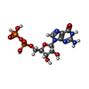
| #7: Chemical | ChemComp-GDP / 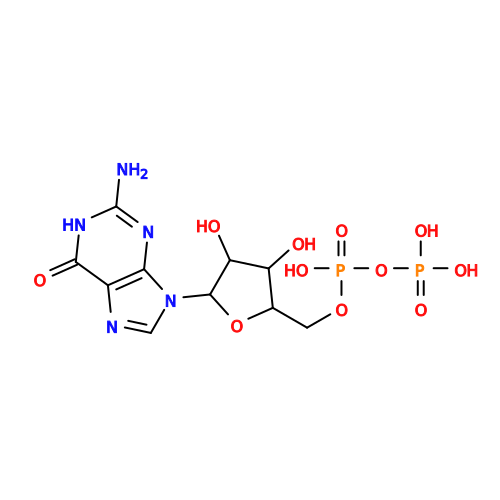 Type: RNA linking / Mass: 443.201 Da / Num. of mol.: 1 / Source method: obtained synthetically / Formula: C10H15N5O11P2 / Feature type: SUBJECT OF INVESTIGATION / Comment: GDP, energy-carrying molecule*YM Type: RNA linking / Mass: 443.201 Da / Num. of mol.: 1 / Source method: obtained synthetically / Formula: C10H15N5O11P2 / Feature type: SUBJECT OF INVESTIGATION / Comment: GDP, energy-carrying molecule*YM |
|---|---|
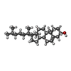
| #8: Chemical | 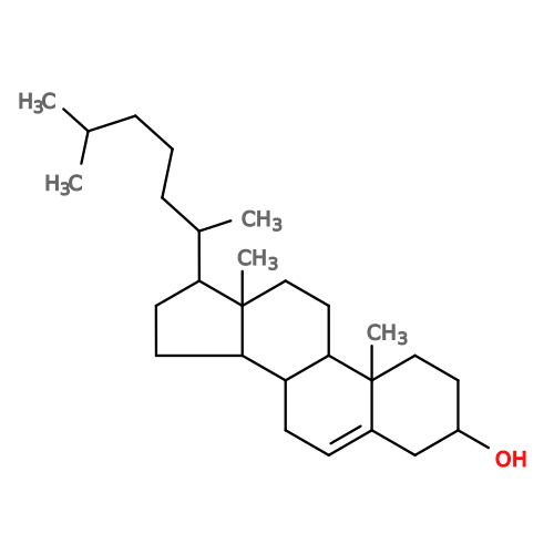 Mass: 386.654 Da / Num. of mol.: 2 / Source method: obtained synthetically / Formula: C27H46O Mass: 386.654 Da / Num. of mol.: 2 / Source method: obtained synthetically / Formula: C27H46O |
-Details
| Has ligand of interest | Y |
|---|---|
| Has protein modification | Y |
-Experimental details
-Experiment
| Experiment | Method: ELECTRON MICROSCOPY |
|---|---|
| EM experiment | Aggregation state: PARTICLE / 3D reconstruction method: single particle reconstruction |
- Sample preparation
| Component |
| |||||||||||||||||||||||||||||||||||
|---|---|---|---|---|---|---|---|---|---|---|---|---|---|---|---|---|---|---|---|---|---|---|---|---|---|---|---|---|---|---|---|---|---|---|---|---|
| Molecular weight |
| |||||||||||||||||||||||||||||||||||
| Source (natural) |
| |||||||||||||||||||||||||||||||||||
| Source (recombinant) |
| |||||||||||||||||||||||||||||||||||
| Buffer solution | pH: 7.2 | |||||||||||||||||||||||||||||||||||
| Buffer component |
| |||||||||||||||||||||||||||||||||||
| Specimen | Conc.: 20 mg/ml / Embedding applied: NO / Shadowing applied: NO / Staining applied: NO / Vitrification applied: YES / Details: chemokine-GPCR complex coupled to Gq heterotrimer | |||||||||||||||||||||||||||||||||||
| Vitrification | Instrument: LEICA EM GP / Cryogen name: ETHANE / Humidity: 95 % / Chamber temperature: 289 K / Details: 3 s blotting before plunging |
- Electron microscopy imaging
| Experimental equipment |  Model: Titan Krios / Image courtesy: FEI Company |
|---|---|
| Microscopy | Model: TFS KRIOS |
| Electron gun | Electron source:  FIELD EMISSION GUN / Accelerating voltage: 300 kV / Illumination mode: FLOOD BEAM FIELD EMISSION GUN / Accelerating voltage: 300 kV / Illumination mode: FLOOD BEAM |
| Electron lens | Mode: BRIGHT FIELD / Nominal magnification: 105000 X / Calibrated magnification: 57624 X / Nominal defocus max: 2000 nm / Nominal defocus min: 800 nm / Cs: 2.7 mm / C2 aperture diameter: 50 µm / Alignment procedure: COMA FREE |
| Specimen holder | Cryogen: NITROGEN / Specimen holder model: FEI TITAN KRIOS AUTOGRID HOLDER |
| Image recording | Average exposure time: 2.5 sec. / Electron dose: 50 e/Å2 / Film or detector model: GATAN K3 (6k x 4k) / Num. of grids imaged: 2 / Num. of real images: 11773 |
| EM imaging optics | Energyfilter name: GIF Bioquantum / Energyfilter slit width: 20 eV |
- Processing
| EM software |
| ||||||||||||||||||||||||||||||||||||||||||||||||||||||||
|---|---|---|---|---|---|---|---|---|---|---|---|---|---|---|---|---|---|---|---|---|---|---|---|---|---|---|---|---|---|---|---|---|---|---|---|---|---|---|---|---|---|---|---|---|---|---|---|---|---|---|---|---|---|---|---|---|---|
| CTF correction | Details: CryoSPARC patch CTF estimation and global/local CTF refinements Type: PHASE FLIPPING AND AMPLITUDE CORRECTION | ||||||||||||||||||||||||||||||||||||||||||||||||||||||||
| Particle selection | Num. of particles selected: 8400483 / Details: initial particles that appeared to be protein | ||||||||||||||||||||||||||||||||||||||||||||||||||||||||
| Symmetry | Point symmetry: C1 (asymmetric) | ||||||||||||||||||||||||||||||||||||||||||||||||||||||||
| 3D reconstruction | Resolution: 2.74 Å / Resolution method: FSC 0.143 CUT-OFF / Num. of particles: 64658 / Algorithm: FOURIER SPACE / Num. of class averages: 1 / Symmetry type: POINT | ||||||||||||||||||||||||||||||||||||||||||||||||||||||||
| Atomic model building | Protocol: FLEXIBLE FIT / Space: REAL | ||||||||||||||||||||||||||||||||||||||||||||||||||||||||
| Atomic model building | 3D fitting-ID: 1
| ||||||||||||||||||||||||||||||||||||||||||||||||||||||||
| Refinement | Cross valid method: NONE Stereochemistry target values: GeoStd + Monomer Library + CDL v1.2 | ||||||||||||||||||||||||||||||||||||||||||||||||||||||||
| Displacement parameters | Biso mean: 44.25 Å2 | ||||||||||||||||||||||||||||||||||||||||||||||||||||||||
| Refine LS restraints |
|