Movie
Movie Controller

[English] 日本語
 Yorodumi
- PDB-9wme: Cryo-EM structure of Clostridium perfringens pili CppA in complex... -
+ Open data

- Basic information
| Entry | Database: PDB / ID: 9wme | |||||||||
|---|---|---|---|---|---|---|---|---|---|---|
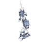
| Title | Cryo-EM structure of Clostridium perfringens pili CppA in complex with CppB | |||||||||
 Components Components |
| |||||||||
 Keywords Keywords | PROTEIN FIBRIL / Shaft pilin / Tip pilin / Clostridium perfringens / surface protein / collagen binding protein | |||||||||
| Function / homology |  Function and homology information Function and homology information | |||||||||
| Biological species |  Clostridium perfringens str. 13 (bacteria) Clostridium perfringens str. 13 (bacteria) | |||||||||
| Method | ELECTRON MICROSCOPY / single particle reconstruction / cryo EM / Resolution: 3.2 Å | |||||||||
 Authors Authors | Nonaka, Y. / Tamai, E. / Kamitori, S. | |||||||||
| Funding support |  Japan, 1items Japan, 1items
| |||||||||
 Citation Citation | Journal: FEBS J / Year: 2026 Title: Dynamic motion of bacterial surface pili based on structural analyses of covalently linked complexes formed by tip and shaft pili proteins from Clostridium perfringens. Authors: Yasuhiro Nonaka / Eiji Tamai / Hiroshi Sekiya / Shigehiro Kamitori / Abstract: The pathogenesis and infectivity of Gram-positive bacteria are mediated by many surface proteins covalently attached to the bacterial cell wall. Pili are types of surface appendages that play ...The pathogenesis and infectivity of Gram-positive bacteria are mediated by many surface proteins covalently attached to the bacterial cell wall. Pili are types of surface appendages that play important roles in the initial adhesion of bacterial cells to host tissues and bacterial colonization. The Gram-positive bacterium Clostridium perfringens (C. perfringens), one of the pathogenic clostridial species causing gas gangrene and food poisoning, has sortase-mediated pili composed of shaft/major pilin A (CppA) and tip/minor pilin B (CppB). The pilus shaft is formed by covalent polymerization of CppA, and CppB is covalently attached to the tip of the shaft involved in adhesion to the host cell. The formation of covalent bonds between CppB and CppA, as well as between CppA and CppA, is catalyzed by class C sortase (CpSrtC), a member of the cysteine transpeptidase family. Since pili consistently have CppB at their tip, CpSrtC is the enzyme that preferentially catalyzes the attachment of CppB (tip) to CppA (shaft) rather than polymerization of CppAs by an unknown mechanism. We determined the structures of complexes formed by covalently linking CppB and CppA by X-ray crystallography and cryo-EM analysis. The complexes have an elongated structure in which β-sandwich folded domains are sequentially arranged, and an amide bond between Thr688 of CppB and Lys174 of CppA was clearly identified. The determined structures allowed us to construct a three-dimensional structure model with dynamic motion of C. perfringens pili, and proposed new insights into the mechanism by which CppB is preferentially attached to CppA. | |||||||||
| History |
|
- Structure visualization
| Structure viewer | Molecule: MolmilJmol/JSmol |
|---|
- Downloads & links
-Download
| PDBx/mmCIF format | 9wme.cif.gz | 134.6 KB | Display | PDBx/mmCIF format |
|---|---|---|---|---|
| PDB format | pdb9wme.ent.gz | 76.5 KB | Display | PDB format |
| PDBx/mmJSON format | 9wme.json.gz | Tree view | PDBx/mmJSON format | |
| Others |  Other downloads Other downloads |
-Validation report
| Arichive directory | https://data.pdbj.org/pub/pdb/validation_reports/wm/9wmeftp://data.pdbj.org/pub/pdb/validation_reports/wm/9wme | HTTPS FTP |
|---|
-Related structure data
| Related structure data |  66088MC  9vo4C  9vo5C M: map data used to model this data C: citing same article ( |
|---|---|
| Similar structure data |
-Links
 PDBj
PDBj- Assembly
| Deposited unit | 
|
|---|---|
| 1 |
|
-Components
| #1: Protein | Mass: 49983.320 Da / Num. of mol.: 1 Source method: isolated from a genetically manipulated source Source: (gene. exp.) Clostridium perfringens str. 13 (bacteria)Gene: CPE0156 / Production host:  Escherichia coli (E. coli) / References: UniProt: Q8XP10 Escherichia coli (E. coli) / References: UniProt: Q8XP10 |
|---|---|
| #2: Protein | Mass: 74482.711 Da / Num. of mol.: 1 Source method: isolated from a genetically manipulated source Source: (gene. exp.) Clostridium perfringens str. 13 (bacteria)Gene: CPE0155 / Production host: Escherichia coli (E. coli) / References: UniProt: Q8XP11 |
| Has protein modification | N |
-Experimental details
-Experiment
| Experiment | Method: ELECTRON MICROSCOPY |
|---|---|
| EM experiment | Aggregation state: PARTICLE / 3D reconstruction method: single particle reconstruction |
- Sample preparation
| Component | Name: Covalent complex of Clostridium perfringens pili proteins CppB and CppA Type: ORGANELLE OR CELLULAR COMPONENT / Entity ID: all / Source: RECOMBINANT |
|---|---|
| Source (natural) | Organism: Clostridium perfringens str. 13 (bacteria) |
| Source (recombinant) | Organism: Escherichia coli (E. coli) |
| Buffer solution | pH: 7.5 / Details: 20 mM Tris-HCl, 100 mM NaCl |
| Specimen | Conc.: 0.26 mg/ml / Embedding applied: NO / Shadowing applied: NO / Staining applied: NO / Vitrification applied: YES |
| Specimen support | Grid material: COPPER / Grid type: Quantifoil R1.2/1.3 |
| Vitrification | Instrument: FEI VITROBOT MARK IV / Cryogen name: ETHANE / Humidity: 100 % / Chamber temperature: 291 K |
- Electron microscopy imaging
| Experimental equipment | 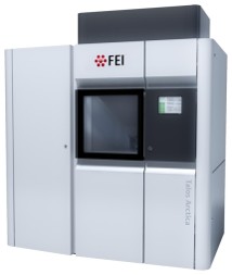 Model: Talos Arctica / Image courtesy: FEI Company |
|---|---|
| Microscopy | Model: FEI TALOS ARCTICA |
| Electron gun | Electron source:  FIELD EMISSION GUN / Accelerating voltage: 200 kV / Illumination mode: FLOOD BEAM FIELD EMISSION GUN / Accelerating voltage: 200 kV / Illumination mode: FLOOD BEAM |
| Electron lens | Mode: BRIGHT FIELD / Nominal defocus max: 2000 nm / Nominal defocus min: 800 nm |
| Image recording | Average exposure time: 3.88 sec. / Electron dose: 50 e/Å2 / Film or detector model: FEI FALCON IV (4k x 4k) / Num. of real images: 3806 |
- Processing
| EM software |
| ||||||||||||||||||||||||||||||||
|---|---|---|---|---|---|---|---|---|---|---|---|---|---|---|---|---|---|---|---|---|---|---|---|---|---|---|---|---|---|---|---|---|---|
| CTF correction | Type: PHASE FLIPPING AND AMPLITUDE CORRECTION | ||||||||||||||||||||||||||||||||
| 3D reconstruction | Resolution: 3.2 Å / Resolution method: FSC 0.143 CUT-OFF / Num. of particles: 103291 / Symmetry type: POINT | ||||||||||||||||||||||||||||||||
| Atomic model building | Protocol: FLEXIBLE FIT / Space: REAL | ||||||||||||||||||||||||||||||||
| Atomic model building | PDB-ID: 5XCC Pdb chain-ID: A / Accession code: 5XCC / Source name: PDB / Type: experimental model | ||||||||||||||||||||||||||||||||
| Refinement | Cross valid method: NONE Stereochemistry target values: GeoStd + Monomer Library + CDL v1.2 | ||||||||||||||||||||||||||||||||
| Displacement parameters | Biso mean: 55.28 Å2 | ||||||||||||||||||||||||||||||||
| Refine LS restraints |
|