 Movie
Movie Controller

[English] 日本語
 Yorodumi
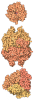
- PDB-9wfz: Cryo-EM structure of PSII PsbA3-S264V from Thermosynechococcus ve... -
+ Open data

- Basic information
| Entry | Database: PDB / ID: 9wfz | |||||||||
|---|---|---|---|---|---|---|---|---|---|---|
| Title | Cryo-EM structure of PSII PsbA3-S264V from Thermosynechococcus vestitus BP-1 (local refinement) | |||||||||
 Components Components |
| |||||||||
 Keywords Keywords | PHOTOSYNTHESIS / PSII mutation / local refinement | |||||||||
| Function / homology |  Function and homology information Function and homology informationoxidoreductase activity, acting on diphenols and related substances as donors, oxygen as acceptor / photosystem II oxygen evolving complex / photosystem II assembly / response to herbicide / oxygen evolving activity / photosystem II stabilization / photosystem II reaction center / photosystem II / photosynthetic electron transport chain / photosystem II ...oxidoreductase activity, acting on diphenols and related substances as donors, oxygen as acceptor / photosystem II oxygen evolving complex / photosystem II assembly / response to herbicide / oxygen evolving activity / photosystem II stabilization / photosystem II reaction center / photosystem II / photosynthetic electron transport chain / photosystem II / extrinsic component of membrane / photosynthetic electron transport in photosystem II / chlorophyll binding / plasma membrane-derived thylakoid membrane / photosynthesis, light reaction / photosynthesis / respiratory electron transport chain / electron transfer activity / iron ion binding / heme binding Similarity search - Function | |||||||||
| Biological species |   Thermosynechococcus vestitus BP-1 (bacteria) Thermosynechococcus vestitus BP-1 (bacteria) | |||||||||
| Method | ELECTRON MICROSCOPY / single particle reconstruction / cryo EM / Resolution: 2.08 Å | |||||||||
 Authors Authors | Fan, S.B. / Nakajima, Y. / Shen, J.R. | |||||||||
| Funding support | 1items
| |||||||||
 Citation Citation | Journal: Biochim Biophys Acta Bioenerg / Year: 2026 Title: Structural and functional analysis of a photosystem II mutant PsbA3-S264V. Authors: Songbo Fan / Yoshiki Nakajima / Koji Kato / Haowei Jiang / Pi-Cheng Tsai / Anqi Jia / Miwa Sugiura / Jian-Ren Shen /  Abstract: Photosystem II (PSII) catalyzes water oxidation and oxygen evolution by a light-induced electron transfer chain, leading to the generation of electrons, protons and dioxygen. D1-S264 is a residue ...Photosystem II (PSII) catalyzes water oxidation and oxygen evolution by a light-induced electron transfer chain, leading to the generation of electrons, protons and dioxygen. D1-S264 is a residue located close to the Q-binding site, and mutation of this residue has been shown to bring significant effects on the electron transfer and oxygen-evolving activities. Here we analyzed the structure of a Thermosynechococcus elongatus mutant PsbA3-S264V by cryo-electron microscopy at 1.96 Å resolution, which showed significant changes in the structure surrounding the bicarbonate and Q-binding region. Due to change of Ser to Val, the hydrogen-bond between the Q carbonyl oxygen and S264 is altered, which changed the protonation pathway of Q from the original route of D1-H252 through D1-S264 to Q, to a new, longer and less efficient route of D1-H252 through D1-F265 to Q. Two residues, D1-E244 and D2-E242, changed their side chain orientations significantly. Among them, D2-E242 adopted two conformations, and both are largely deviated from the original structure. All these changes led to alterations in hydrogen-bonding networks of two channels, channel A and channel B, that connect the stromal surface to Q and may function to transport protons to protonate Q. Furthermore, isothermal titration calorimetry experiments showed a diminished 3-(3,4-dichlorophenyl)-1, 1-dimethylurea (DCMU) binding affinity of the mutated PSII, which may be explained by a structural rotation of D1-F255 in the mutant based on structural analysis of DCMU-bound PSII. These findings offer valuable insights into the functions of D1-S264 in Q protonation and function, as well as in the DCMU-binding. | |||||||||
| History |
|
- Structure visualization
| Structure viewer | Molecule: MolmilJmol/JSmol |
|---|
- Downloads & links
-Download
| PDBx/mmCIF format | 9wfz.cif.gz | 506.5 KB | Display | PDBx/mmCIF format |
|---|---|---|---|---|
| PDB format | pdb9wfz.ent.gz | 377.2 KB | Display | PDB format |
| PDBx/mmJSON format | 9wfz.json.gz | Tree view | PDBx/mmJSON format | |
| Others |  Other downloads Other downloads |
-Validation report
| Arichive directory | https://data.pdbj.org/pub/pdb/validation_reports/wf/9wfzftp://data.pdbj.org/pub/pdb/validation_reports/wf/9wfz | HTTPS FTP |
|---|
-Related structure data
| Related structure data |  65941MC  9w5bC  9w7dC M: map data used to model this data C: citing same article ( |
|---|---|
| Similar structure data |


-Links
 PDBj
PDBj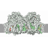
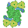
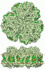
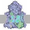

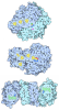


- Assembly
| Deposited unit | 
|
|---|---|
| 1 |
|
-Components
-Photosystem II ... , 16 types, 17 molecules IKTVYABbCDJLMOUXZ
| #1: Protein/peptide | Mass: 4410.245 Da / Num. of mol.: 1 / Source method: isolated from a natural source Source: (natural) Thermosynechococcus vestitus BP-1 (bacteria)References: UniProt: Q8DJZ6 | ||||||||||||||||||
|---|---|---|---|---|---|---|---|---|---|---|---|---|---|---|---|---|---|---|---|
| #2: Protein/peptide | Mass: 5028.083 Da / Num. of mol.: 1 / Source method: isolated from a natural source Source: (natural) Thermosynechococcus vestitus BP-1 (bacteria)References: UniProt: Q9F1K9 | ||||||||||||||||||
| #3: Protein/peptide | Mass: 3878.728 Da / Num. of mol.: 1 / Source method: isolated from a natural source Source: (natural) Thermosynechococcus vestitus BP-1 (bacteria)References: UniProt: Q8DIQ0 | ||||||||||||||||||
| #4: Protein | Mass: 18046.943 Da / Num. of mol.: 1 / Source method: isolated from a natural source Source: (natural) Thermosynechococcus vestitus BP-1 (bacteria)References: UniProt: P0A386 | ||||||||||||||||||
| #5: Protein/peptide | Mass: 5039.143 Da / Num. of mol.: 1 / Source method: isolated from a natural source Source: (natural) Thermosynechococcus vestitus BP-1 (bacteria)References: UniProt: Q8DJI1 | ||||||||||||||||||
| #6: Protein | Mass: 39792.391 Da / Num. of mol.: 1 / Source method: isolated from a natural source Source: (natural) Thermosynechococcus vestitus BP-1 (bacteria)References: UniProt: Q8DIV4, photosystem II | ||||||||||||||||||
| #7: Protein | Mass: 56656.457 Da / Num. of mol.: 2 / Source method: isolated from a natural source Source: (natural) Thermosynechococcus vestitus BP-1 (bacteria)References: UniProt: Q8DIQ1 #8: Protein | | Mass: 50287.500 Da / Num. of mol.: 1 / Source method: isolated from a natural source Source: (natural) Thermosynechococcus vestitus BP-1 (bacteria)References: UniProt: Q8DIF8 #9: Protein | | Mass: 39388.156 Da / Num. of mol.: 1 / Source method: isolated from a natural source Source: (natural) Thermosynechococcus vestitus BP-1 (bacteria)References: UniProt: Q8CM25, photosystem II #12: Protein/peptide | | Mass: 4105.908 Da / Num. of mol.: 1 / Source method: isolated from a natural source Source: (natural) Thermosynechococcus vestitus BP-1 (bacteria)References: UniProt: P59087 #13: Protein/peptide | | Mass: 4299.044 Da / Num. of mol.: 1 / Source method: isolated from a natural source Source: (natural) Thermosynechococcus vestitus BP-1 (bacteria)References: UniProt: Q8DIN8 #14: Protein/peptide | | Mass: 3981.673 Da / Num. of mol.: 1 / Source method: isolated from a natural source Source: (natural) Thermosynechococcus vestitus BP-1 (bacteria)References: UniProt: Q8DHA7 #15: Protein | | Mass: 29637.443 Da / Num. of mol.: 1 / Source method: isolated from a natural source Source: (natural) Thermosynechococcus vestitus BP-1 (bacteria)References: UniProt: P0A431 #16: Protein | | Mass: 15030.986 Da / Num. of mol.: 1 / Source method: isolated from a natural source Source: (natural) Thermosynechococcus vestitus BP-1 (bacteria)References: UniProt: Q9F1L5 #17: Protein/peptide | | Mass: 4322.226 Da / Num. of mol.: 1 / Source method: isolated from a natural source Source: (natural) Thermosynechococcus vestitus BP-1 (bacteria)References: UniProt: Q9F1R6 #18: Protein | | Mass: 6766.187 Da / Num. of mol.: 1 / Source method: isolated from a natural source Source: (natural) Thermosynechococcus vestitus BP-1 (bacteria)References: UniProt: Q8DHJ2 |
-Cytochrome b559 subunit ... , 2 types, 2 molecules EF
| #10: Protein | Mass: 9580.840 Da / Num. of mol.: 1 / Source method: isolated from a natural source Source: (natural) Thermosynechococcus vestitus BP-1 (bacteria)References: UniProt: Q8DIP0 |
|---|---|
| #11: Protein/peptide | Mass: 5067.900 Da / Num. of mol.: 1 / Source method: isolated from a natural source Source: (natural) Thermosynechococcus vestitus BP-1 (bacteria)References: UniProt: Q8DIN9 |
-Sugars , 2 types, 7 molecules 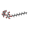
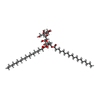
| #21: Sugar | 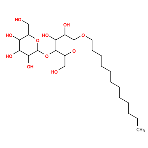 Type: D-saccharide / Mass: 510.615 Da / Num. of mol.: 3 / Source method: obtained synthetically / Formula: C24H46O11 / Comment: detergent*YM Type: D-saccharide / Mass: 510.615 Da / Num. of mol.: 3 / Source method: obtained synthetically / Formula: C24H46O11 / Comment: detergent*YM#33: Sugar | ChemComp-DGD /  Type: saccharide / Mass: 949.299 Da / Num. of mol.: 4 / Source method: obtained synthetically / Formula: C51H96O15 Type: saccharide / Mass: 949.299 Da / Num. of mol.: 4 / Source method: obtained synthetically / Formula: C51H96O15 |
|---|
-Non-polymers , 15 types, 497 molecules 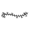
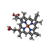
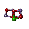


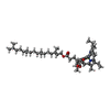

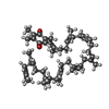
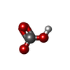
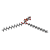
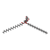
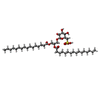
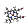

| #19: Chemical | ChemComp-UNL / Num. of mol.: 6 / Source method: obtained synthetically #20: Chemical | ChemComp-BCR / 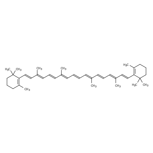 Mass: 536.873 Da / Num. of mol.: 8 / Source method: obtained synthetically / Formula: C40H56 Mass: 536.873 Da / Num. of mol.: 8 / Source method: obtained synthetically / Formula: C40H56#22: Chemical | ChemComp-HEC / |  Mass: 620.519 Da / Num. of mol.: 1 / Source method: obtained synthetically / Formula: C34H36FeN4O4 Mass: 620.519 Da / Num. of mol.: 1 / Source method: obtained synthetically / Formula: C34H36FeN4O4#23: Chemical | ChemComp-OEX / | 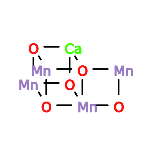 Mass: 339.827 Da / Num. of mol.: 1 / Source method: obtained synthetically / Formula: CaMn4O5 Mass: 339.827 Da / Num. of mol.: 1 / Source method: obtained synthetically / Formula: CaMn4O5#24: Chemical | ChemComp-FE2 / |  Mass: 55.845 Da / Num. of mol.: 1 / Source method: obtained synthetically / Formula: Fe Mass: 55.845 Da / Num. of mol.: 1 / Source method: obtained synthetically / Formula: Fe#25: Chemical |  Mass: 35.453 Da / Num. of mol.: 2 / Source method: obtained synthetically / Formula: Cl Mass: 35.453 Da / Num. of mol.: 2 / Source method: obtained synthetically / Formula: Cl#26: Chemical | ChemComp-CLA / 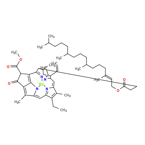 Mass: 893.489 Da / Num. of mol.: 22 / Source method: obtained synthetically / Formula: C55H72MgN4O5 Mass: 893.489 Da / Num. of mol.: 22 / Source method: obtained synthetically / Formula: C55H72MgN4O5#27: Chemical | 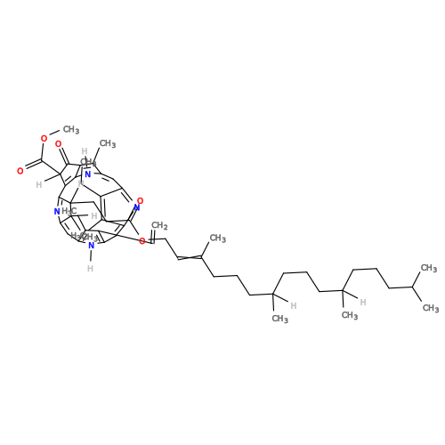 Mass: 871.200 Da / Num. of mol.: 2 / Source method: obtained synthetically / Formula: C55H74N4O5 Mass: 871.200 Da / Num. of mol.: 2 / Source method: obtained synthetically / Formula: C55H74N4O5#28: Chemical | 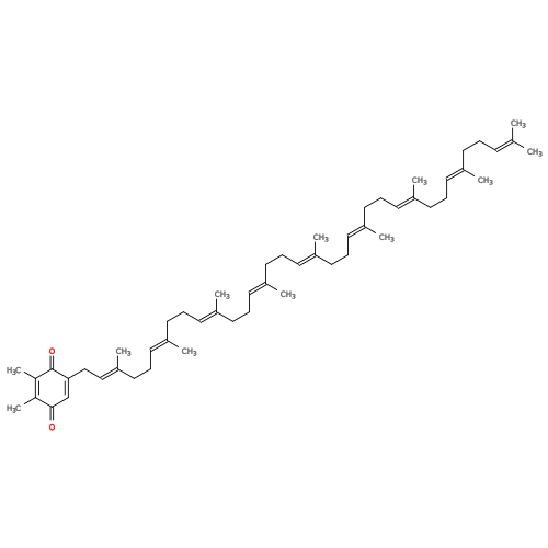 Mass: 749.201 Da / Num. of mol.: 2 / Source method: obtained synthetically / Formula: C53H80O2 Mass: 749.201 Da / Num. of mol.: 2 / Source method: obtained synthetically / Formula: C53H80O2#29: Chemical | ChemComp-BCT / | 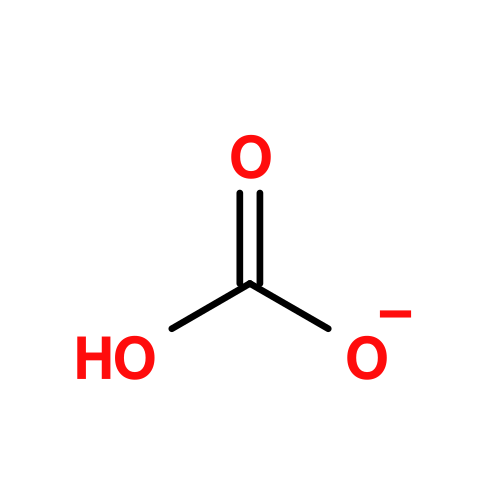 Mass: 61.017 Da / Num. of mol.: 1 / Source method: obtained synthetically / Formula: CHO3 / Comment: pH buffer*YM Mass: 61.017 Da / Num. of mol.: 1 / Source method: obtained synthetically / Formula: CHO3 / Comment: pH buffer*YM#30: Chemical | ChemComp-LHG / 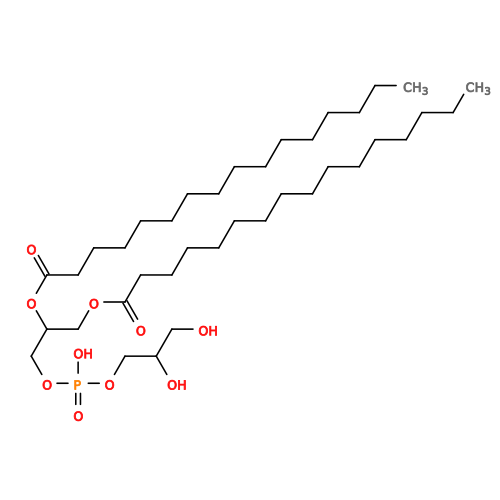 Mass: 722.970 Da / Num. of mol.: 5 / Source method: obtained synthetically / Formula: C38H75O10P / Comment: phospholipid*YM Mass: 722.970 Da / Num. of mol.: 5 / Source method: obtained synthetically / Formula: C38H75O10P / Comment: phospholipid*YM#31: Chemical | 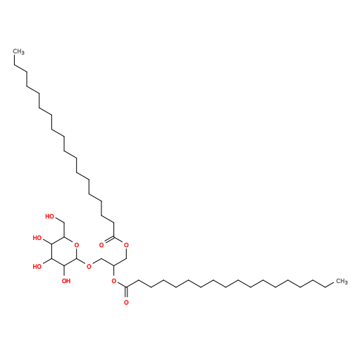 Mass: 787.158 Da / Num. of mol.: 3 / Source method: obtained synthetically / Formula: C45H86O10 Mass: 787.158 Da / Num. of mol.: 3 / Source method: obtained synthetically / Formula: C45H86O10#32: Chemical | 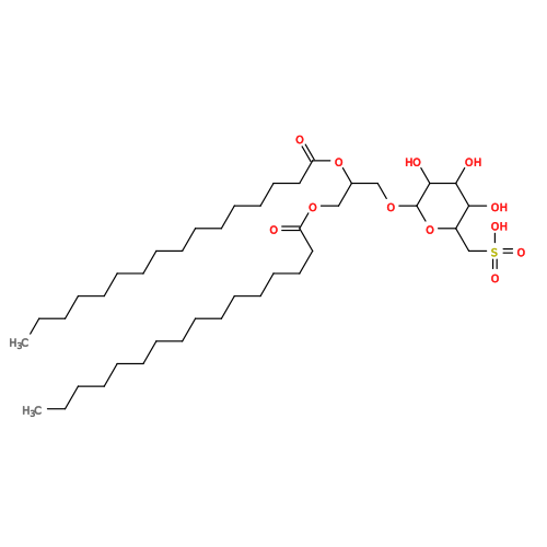 Mass: 795.116 Da / Num. of mol.: 2 / Source method: obtained synthetically / Formula: C41H78O12S Mass: 795.116 Da / Num. of mol.: 2 / Source method: obtained synthetically / Formula: C41H78O12S#34: Chemical | ChemComp-HEM / | 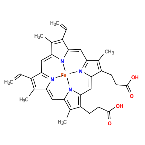 Mass: 616.487 Da / Num. of mol.: 1 / Source method: obtained synthetically / Formula: C34H32FeN4O4 Mass: 616.487 Da / Num. of mol.: 1 / Source method: obtained synthetically / Formula: C34H32FeN4O4#35: Water | ChemComp-HOH / | Mass: 18.015 Da / Num. of mol.: 440 / Source method: isolated from a natural source / Formula: H2O |
|---|
-Details
| Has ligand of interest | N |
|---|---|
| Has protein modification | Y |
-Experimental details
-Experiment
| Experiment | Method: ELECTRON MICROSCOPY |
|---|---|
| EM experiment | Aggregation state: PARTICLE / 3D reconstruction method: single particle reconstruction |
- Sample preparation
| Component | Name: Cryo-EM structure of PSII PsbA3-S264V from Thermosynechococcus vestitus BP-1 (local refinement) Type: COMPLEX / Entity ID: #1-#18 / Source: NATURAL |
|---|---|
| Source (natural) | Organism: Thermosynechococcus vestitus BP-1 (bacteria) |
| Buffer solution | pH: 6.5 |
| Specimen | Embedding applied: NO / Shadowing applied: NO / Staining applied: NO / Vitrification applied: YES |
| Vitrification | Cryogen name: ETHANE / Humidity: 100 % |
- Electron microscopy imaging
| Experimental equipment |  Model: Titan Krios / Image courtesy: FEI Company |
|---|---|
| Microscopy | Model: TFS KRIOS |
| Electron gun | Electron source:  FIELD EMISSION GUN / Accelerating voltage: 300 kV / Illumination mode: FLOOD BEAM FIELD EMISSION GUN / Accelerating voltage: 300 kV / Illumination mode: FLOOD BEAM |
| Electron lens | Mode: BRIGHT FIELD / Nominal defocus max: 1000 nm / Nominal defocus min: 200 nm |
| Image recording | Electron dose: 50 e/Å2 / Film or detector model: FEI FALCON IV (4k x 4k) |
- Processing
| EM software |
| ||||||||||||||||
|---|---|---|---|---|---|---|---|---|---|---|---|---|---|---|---|---|---|
| CTF correction | Type: PHASE FLIPPING AND AMPLITUDE CORRECTION | ||||||||||||||||
| 3D reconstruction | Resolution: 2.08 Å / Resolution method: FSC 0.143 CUT-OFF / Num. of particles: 93573 / Symmetry type: POINT |
