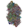
Movie
Movie Controller

[English] 日本語
 Yorodumi
- EMDB-65941: Cryo-EM structure of PSII PsbA3-S264V from Thermosynechococcus ve... -
+ Open data

- Basic information
| Entry |  | |||||||||
|---|---|---|---|---|---|---|---|---|---|---|
| Title | Cryo-EM structure of PSII PsbA3-S264V from Thermosynechococcus vestitus BP-1 (local refinement) | |||||||||
 Map data Map data | ||||||||||
 Sample Sample |
| |||||||||
 Keywords Keywords | PSII mutation / local refinement / PHOTOSYNTHESIS | |||||||||
| Function / homology |  Function and homology information Function and homology informationoxidoreductase activity, acting on diphenols and related substances as donors, oxygen as acceptor / photosystem II oxygen evolving complex / photosystem II assembly / response to herbicide / oxygen evolving activity / photosystem II stabilization / photosystem II reaction center / photosystem II / photosynthetic electron transport chain / photosystem II ...oxidoreductase activity, acting on diphenols and related substances as donors, oxygen as acceptor / photosystem II oxygen evolving complex / photosystem II assembly / response to herbicide / oxygen evolving activity / photosystem II stabilization / photosystem II reaction center / photosystem II / photosynthetic electron transport chain / photosystem II / extrinsic component of membrane / photosynthetic electron transport in photosystem II / chlorophyll binding / plasma membrane-derived thylakoid membrane / photosynthesis, light reaction / photosynthesis / respiratory electron transport chain / electron transfer activity / iron ion binding / heme binding Similarity search - Function | |||||||||
| Biological species |   Thermosynechococcus vestitus BP-1 (bacteria) Thermosynechococcus vestitus BP-1 (bacteria) | |||||||||
| Method | single particle reconstruction / cryo EM / Resolution: 2.08 Å | |||||||||
 Authors Authors | Fan SB / Nakajima Y / Shen JR | |||||||||
| Funding support | 1 items
| |||||||||
 Citation Citation | Journal: Biochim Biophys Acta Bioenerg / Year: 2026 Title: Structural and functional analysis of a photosystem II mutant PsbA3-S264V. Authors: Songbo Fan / Yoshiki Nakajima / Koji Kato / Haowei Jiang / Pi-Cheng Tsai / Anqi Jia / Miwa Sugiura / Jian-Ren Shen /  Abstract: Photosystem II (PSII) catalyzes water oxidation and oxygen evolution by a light-induced electron transfer chain, leading to the generation of electrons, protons and dioxygen. D1-S264 is a residue ...Photosystem II (PSII) catalyzes water oxidation and oxygen evolution by a light-induced electron transfer chain, leading to the generation of electrons, protons and dioxygen. D1-S264 is a residue located close to the Q-binding site, and mutation of this residue has been shown to bring significant effects on the electron transfer and oxygen-evolving activities. Here we analyzed the structure of a Thermosynechococcus elongatus mutant PsbA3-S264V by cryo-electron microscopy at 1.96 Å resolution, which showed significant changes in the structure surrounding the bicarbonate and Q-binding region. Due to change of Ser to Val, the hydrogen-bond between the Q carbonyl oxygen and S264 is altered, which changed the protonation pathway of Q from the original route of D1-H252 through D1-S264 to Q, to a new, longer and less efficient route of D1-H252 through D1-F265 to Q. Two residues, D1-E244 and D2-E242, changed their side chain orientations significantly. Among them, D2-E242 adopted two conformations, and both are largely deviated from the original structure. All these changes led to alterations in hydrogen-bonding networks of two channels, channel A and channel B, that connect the stromal surface to Q and may function to transport protons to protonate Q. Furthermore, isothermal titration calorimetry experiments showed a diminished 3-(3,4-dichlorophenyl)-1, 1-dimethylurea (DCMU) binding affinity of the mutated PSII, which may be explained by a structural rotation of D1-F255 in the mutant based on structural analysis of DCMU-bound PSII. These findings offer valuable insights into the functions of D1-S264 in Q protonation and function, as well as in the DCMU-binding. | |||||||||
| History |
|
- Structure visualization
| Supplemental images |
|---|

- Downloads & links
-EMDB archive
| Map data | emd_65941.map.gz | 483.2 MB | EMDB map data format | |
|---|---|---|---|---|
| Header (meta data) | emd-65941-v30.xmlemd-65941.xml | 37.6 KB 37.6 KB | Display Display | EMDB header |
| Images |  emd_65941.png emd_65941.png | 63.9 KB | ||
| Filedesc metadata | emd-65941.cif.gz | 8.7 KB | ||
| Others | emd_65941_half_map_1.map.gzemd_65941_half_map_2.map.gz | 475.3 MB 475.3 MB | ||
| Archive directory |  https://data.pdbj.org/pub/emdb/structures/EMD-65941ftp://data.pdbj.org/pub/emdb/structures/EMD-65941 https://data.pdbj.org/pub/emdb/structures/EMD-65941ftp://data.pdbj.org/pub/emdb/structures/EMD-65941 | HTTPS FTP |
-Related structure data
| Related structure data |  9wfzMC  9w5bC  9w7dC M: atomic model generated by this map C: citing same article ( |
|---|---|
| Similar structure data |


-Links
| EMDB pages | EMDB (EBI/PDBe) / EMDataResource |
|---|---|
| Related items in Molecule of the Month |
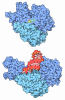
-Map
| File | Download / File: emd_65941.map.gz / Format: CCP4 / Size: 512 MB / Type: IMAGE STORED AS FLOATING POINT NUMBER (4 BYTES) | ||||||||||||||||||||||||||||||||||||
|---|---|---|---|---|---|---|---|---|---|---|---|---|---|---|---|---|---|---|---|---|---|---|---|---|---|---|---|---|---|---|---|---|---|---|---|---|---|
| Projections & slices | Image control
Images are generated by Spider. | ||||||||||||||||||||||||||||||||||||
| Voxel size | X=Y=Z: 0.5675 Å | ||||||||||||||||||||||||||||||||||||
| Density |
| ||||||||||||||||||||||||||||||||||||
| Symmetry | Space group: 1 | ||||||||||||||||||||||||||||||||||||
| Details | EMDB XML:
|
 Z (Sec.)
 Y (Row.)
 X (Col.)


-Supplemental data
-Half map: #2
| File | emd_65941_half_map_1.map | ||||||||||||
|---|---|---|---|---|---|---|---|---|---|---|---|---|---|
| Projections & Slices |
| ||||||||||||
| Density Histograms |


-Half map: #1
| File | emd_65941_half_map_2.map | ||||||||||||
|---|---|---|---|---|---|---|---|---|---|---|---|---|---|
| Projections & Slices |
| ||||||||||||
| Density Histograms |


- Sample components
+Entire : Cryo-EM structure of PSII PsbA3-S264V from Thermosynechococcus ve...
+Supramolecule #1: Cryo-EM structure of PSII PsbA3-S264V from Thermosynechococcus ve...
+Macromolecule #1: Photosystem II reaction center protein I
+Macromolecule #2: Photosystem II reaction center protein K
+Macromolecule #3: Photosystem II reaction center protein T
+Macromolecule #4: Photosystem II extrinsic protein V
+Macromolecule #5: Photosystem II reaction center protein Psb30
+Macromolecule #6: Photosystem II protein D1 3
+Macromolecule #7: Photosystem II CP47 reaction center protein
+Macromolecule #8: Photosystem II CP43 reaction center protein
+Macromolecule #9: Photosystem II D2 protein
+Macromolecule #10: Cytochrome b559 subunit alpha
+Macromolecule #11: Cytochrome b559 subunit beta
+Macromolecule #12: Photosystem II reaction center protein J
+Macromolecule #13: Photosystem II reaction center protein L
+Macromolecule #14: Photosystem II reaction center protein M
+Macromolecule #15: Photosystem II extrinsic protein O
+Macromolecule #16: Photosystem II extrinsic protein U
+Macromolecule #17: Photosystem II reaction center protein X
+Macromolecule #18: Photosystem II reaction center protein Z
+Macromolecule #19: UNKNOWN LIGAND

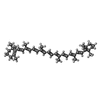
+Macromolecule #20: BETA-CAROTENE
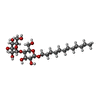
+Macromolecule #21: DODECYL-BETA-D-MALTOSIDE
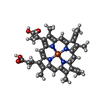
+Macromolecule #22: HEME C
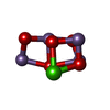
+Macromolecule #23: CA-MN4-O5 CLUSTER
+Macromolecule #24: FE (II) ION
+Macromolecule #25: CHLORIDE ION
+Macromolecule #26: CHLOROPHYLL A
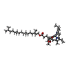
+Macromolecule #27: PHEOPHYTIN A

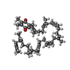
+Macromolecule #28: 2,3-DIMETHYL-5-(3,7,11,15,19,23,27,31,35-NONAMETHYL-2,6,10,14,18,...
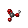
+Macromolecule #29: BICARBONATE ION
+Macromolecule #30: 1,2-DIPALMITOYL-PHOSPHATIDYL-GLYCEROLE
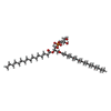
+Macromolecule #31: 1,2-DISTEAROYL-MONOGALACTOSYL-DIGLYCERIDE
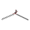
+Macromolecule #32: 1,2-DI-O-ACYL-3-O-[6-DEOXY-6-SULFO-ALPHA-D-GLUCOPYRANOSYL]-SN-GLYCEROL
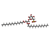
+Macromolecule #33: DIGALACTOSYL DIACYL GLYCEROL (DGDG)
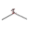
+Macromolecule #34: PROTOPORPHYRIN IX CONTAINING FE
+Macromolecule #35: water

-Experimental details
-Structure determination
| Method | cryo EM |
|---|---|
 Processing Processing | single particle reconstruction |
| Aggregation state | particle |
-Sample preparation
| Buffer | pH: 6.5 |
|---|---|
| Vitrification | Cryogen name: ETHANE / Chamber humidity: 100 % |
- Electron microscopy
| Microscope | TFS KRIOS |
|---|---|
| Image recording | Film or detector model: FEI FALCON IV (4k x 4k) / Average electron dose: 50.0 e/Å2 |
| Electron beam | Acceleration voltage: 300 kV / Electron source:  FIELD EMISSION GUN FIELD EMISSION GUN |
| Electron optics | Illumination mode: FLOOD BEAM / Imaging mode: BRIGHT FIELD / Nominal defocus max: 1.0 µm / Nominal defocus min: 0.2 µm |
| Experimental equipment |  Model: Titan Krios / Image courtesy: FEI Company |