Movie
Movie Controller


+ Open data

- Basic information
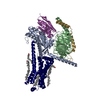
| Entry | Database: PDB / ID: 9wey | ||||||||||||||||||||||||||||||||||||
|---|---|---|---|---|---|---|---|---|---|---|---|---|---|---|---|---|---|---|---|---|---|---|---|---|---|---|---|---|---|---|---|---|---|---|---|---|---|
| Title | Structure of HCMV UL33 in complex with human Gs protein | ||||||||||||||||||||||||||||||||||||
 Components Components |
| ||||||||||||||||||||||||||||||||||||
 Keywords Keywords | MEMBRANE PROTEIN / HCMV / viral protein / viral-host interaction / GPCR | ||||||||||||||||||||||||||||||||||||
| Function / homology |  Function and homology information Function and homology informationadenylate cyclase-activating G protein-coupled bile acid receptor signaling pathway / adenylate cyclase-activating serotonin receptor signaling pathway / regulation of skeletal muscle contraction / PKA activation in glucagon signalling / hair follicle placode formation / developmental growth / intracellular transport / D1 dopamine receptor binding / vascular endothelial cell response to laminar fluid shear stress / renal water homeostasis ...adenylate cyclase-activating G protein-coupled bile acid receptor signaling pathway / adenylate cyclase-activating serotonin receptor signaling pathway / regulation of skeletal muscle contraction / PKA activation in glucagon signalling / hair follicle placode formation / developmental growth / intracellular transport / D1 dopamine receptor binding / vascular endothelial cell response to laminar fluid shear stress / renal water homeostasis / Hedgehog 'off' state / activation of adenylate cyclase activity / adenylate cyclase-activating adrenergic receptor signaling pathway / cellular response to acidic pH / cellular response to glucagon stimulus / intracellular glucose homeostasis / positive regulation of insulin secretion involved in cellular response to glucose stimulus / adenylate cyclase activator activity / trans-Golgi network membrane / negative regulation of inflammatory response to antigenic stimulus / response to prostaglandin E / bone development / platelet aggregation / cognition / G-protein beta/gamma-subunit complex binding / positive regulation of insulin secretion / Olfactory Signaling Pathway / Activation of the phototransduction cascade / G protein-coupled acetylcholine receptor signaling pathway / G beta:gamma signalling through PLC beta / Presynaptic function of Kainate receptors / Thromboxane signalling through TP receptor / sensory perception of smell / Activation of G protein gated Potassium channels / Inhibition of voltage gated Ca2+ channels via Gbeta/gamma subunits / G-protein activation / Glucagon signaling in metabolic regulation / G beta:gamma signalling through CDC42 / Prostacyclin signalling through prostacyclin receptor / Synthesis, secretion, and inactivation of Glucagon-like Peptide-1 (GLP-1) / G beta:gamma signalling through BTK / photoreceptor disc membrane / ADP signalling through P2Y purinoceptor 12 / Glucagon-type ligand receptors / Sensory perception of sweet, bitter, and umami (glutamate) taste / Adrenaline,noradrenaline inhibits insulin secretion / Vasopressin regulates renal water homeostasis via Aquaporins / Glucagon-like Peptide-1 (GLP1) regulates insulin secretion / G alpha (z) signalling events / cellular response to catecholamine stimulus / ADP signalling through P2Y purinoceptor 1 / G beta:gamma signalling through PI3Kgamma / ADORA2B mediated anti-inflammatory cytokines production / adenylate cyclase-activating dopamine receptor signaling pathway / Cooperation of PDCL (PhLP1) and TRiC/CCT in G-protein beta folding / positive regulation of cold-induced thermogenesis / GPER1 signaling / cellular response to prostaglandin E stimulus / heterotrimeric G-protein complex / Inactivation, recovery and regulation of the phototransduction cascade / G alpha (12/13) signalling events / G-protein beta-subunit binding / extracellular vesicle / sensory perception of taste / Thrombin signalling through proteinase activated receptors (PARs) / signaling receptor complex adaptor activity / adenylate cyclase-activating G protein-coupled receptor signaling pathway / retina development in camera-type eye / fibroblast proliferation / GTPase binding / G protein activity / Ca2+ pathway / High laminar flow shear stress activates signaling by PIEZO1 and PECAM1:CDH5:KDR in endothelial cells / G alpha (i) signalling events / G alpha (s) signalling events / G alpha (q) signalling events / phospholipase C-activating G protein-coupled receptor signaling pathway / Hydrolases; Acting on acid anhydrides; Acting on GTP to facilitate cellular and subcellular movement / Ras protein signal transduction / cell population proliferation / Extra-nuclear estrogen signaling / G protein-coupled receptor signaling pathway / lysosomal membrane / GTPase activity / synapse / GTP binding / protein-containing complex binding / signal transduction / extracellular exosome / membrane / metal ion binding / plasma membrane / cytosol / cytoplasm Similarity search - Function | ||||||||||||||||||||||||||||||||||||
| Biological species |  Homo sapiens (human) Homo sapiens (human)   Human betaherpesvirus 5 Human betaherpesvirus 5 | ||||||||||||||||||||||||||||||||||||
| Method | ELECTRON MICROSCOPY / single particle reconstruction / cryo EM / Resolution: 3.3 Å | ||||||||||||||||||||||||||||||||||||
 Authors Authors | Tsutsumi, N. / Suzuki, S. / Nishikawa, K. / Fujiyoshi, Y. | ||||||||||||||||||||||||||||||||||||
| Funding support |  Japan, 1items Japan, 1items
| ||||||||||||||||||||||||||||||||||||
 Citation Citation | Journal: Commun Biol / Year: 2026 Title: Activation of cytomegalovirus-encoded G protein-coupled receptor UL33 by an innate N-terminal peptide. Authors: Anna K Drzazga / Shota Suzuki / Caroline Wouters / Felix Faas / Kouki Nishikawa / Akiko Kamegawa / Yoshinori Fujiyoshi / Mette M Rosenkilde / Naotaka Tsutsumi /  Abstract: Human cytomegalovirus (HCMV) encodes the orphan G protein-coupled receptor (GPCR) UL33, which exhibits constitutive activity that disrupts host G protein signalling, facilitating efficient viral ...Human cytomegalovirus (HCMV) encodes the orphan G protein-coupled receptor (GPCR) UL33, which exhibits constitutive activity that disrupts host G protein signalling, facilitating efficient viral replication and pathogenesis. The cryo-electron microscopy (cryo-EM) structure of UL33 bound to the G subtype of G protein reveals the N-terminal peptide as a tethered ligand reminiscent of the protease-activated receptors and adhesion GPCRs. This self-agonism induces a non-canonical active state that facilitates promiscuous G protein coupling, a plausible viral strategy for fine-tuning host signalling. Structure-guided mutagenesis disrupting key interactions between the N-terminus and its binding pocket abolishes G protein-mediated signalling, confirming the role of the N-terminus as a self-agonist. Our findings elucidate the structural basis for this activation mechanism and highlight the strategies employed by HCMV to hijack host G protein signalling. | ||||||||||||||||||||||||||||||||||||
| History |
|
- Structure visualization
| Structure viewer | Molecule: MolmilJmol/JSmol |
|---|
- Downloads & links
-Download
| PDBx/mmCIF format | 9wey.cif.gz | 220.6 KB | Display | PDBx/mmCIF format |
|---|---|---|---|---|
| PDB format | pdb9wey.ent.gz | 164.4 KB | Display | PDB format |
| PDBx/mmJSON format | 9wey.json.gz | Tree view | PDBx/mmJSON format | |
| Others |  Other downloads Other downloads |
-Validation report
| Arichive directory | https://data.pdbj.org/pub/pdb/validation_reports/we/9weyftp://data.pdbj.org/pub/pdb/validation_reports/we/9wey | HTTPS FTP |
|---|
-Related structure data
| Related structure data |  65918MC M: map data used to model this data C: citing same article ( |
|---|---|
| Similar structure data |
-Links
 PDBj
PDBj


- Assembly
| Deposited unit | 
|
|---|---|
| 1 |
|
-Components
| #1: Protein | Mass: 7729.947 Da / Num. of mol.: 1 Source method: isolated from a genetically manipulated source Source: (gene. exp.) Homo sapiens (human) / Gene: GNG2 / Production host: Homo sapiens (human) / References: UniProt: P59768 |
|---|---|
| #2: Antibody | Mass: 15969.623 Da / Num. of mol.: 1 Source method: isolated from a genetically manipulated source Source: (gene. exp.)  |
| #3: Protein | Mass: 45568.238 Da / Num. of mol.: 1 Mutation: S54N, G226A, E268A, N271K, K274D, R280K, T284D, I285T, A366S Source method: isolated from a genetically manipulated source Source: (gene. exp.) Homo sapiens (human) / Gene: GNAS, GNAS1, GSP / Production host: Homo sapiens (human)References: UniProt: P63092, Hydrolases; Acting on acid anhydrides; Acting on GTP to facilitate cellular and subcellular movement |
| #4: Protein | Mass: 40039.648 Da / Num. of mol.: 1 Source method: isolated from a genetically manipulated source Source: (gene. exp.) Homo sapiens (human) / Gene: GNB1 / Production host: Homo sapiens (human) / References: UniProt: P62873 |
| #5: Protein | Mass: 68188.695 Da / Num. of mol.: 1 Source method: isolated from a genetically manipulated source Details: G-protein coupled receptor homolog UL33, LgBit fusion,G-protein coupled receptor homolog UL33, LgBit fusion,G-protein coupled receptor homolog UL33, LgBit fusion,G-protein coupled receptor ...Details: G-protein coupled receptor homolog UL33, LgBit fusion,G-protein coupled receptor homolog UL33, LgBit fusion,G-protein coupled receptor homolog UL33, LgBit fusion,G-protein coupled receptor homolog UL33, LgBit fusion Source: (gene. exp.) Human betaherpesvirus 5 / Gene: UL33 / Production host: Human betaherpesvirus 5 |
| Has protein modification | Y |
-Experimental details
-Experiment
| Experiment | Method: ELECTRON MICROSCOPY |
|---|---|
| EM experiment | Aggregation state: PARTICLE / 3D reconstruction method: single particle reconstruction |
- Sample preparation
| Component | Name: UL33-Gas-Gb1-Gg2-Nb35 / Type: COMPLEX Details: Cytomegalovirus GPCR UL33 complexed with heterotrimeric Gs protein with dominant negative mutations via HiBit tethering stabilized by nanobody35 Entity ID: all / Source: RECOMBINANT | ||||||||||||||||||||||||
|---|---|---|---|---|---|---|---|---|---|---|---|---|---|---|---|---|---|---|---|---|---|---|---|---|---|
| Molecular weight | Value: 0.18 MDa / Experimental value: NO | ||||||||||||||||||||||||
| Source (natural) | Organism: Homo sapiens (human) | ||||||||||||||||||||||||
| Source (recombinant) | Organism: Homo sapiens (human) | ||||||||||||||||||||||||
| Buffer solution | pH: 7.2 | ||||||||||||||||||||||||
| Buffer component |
| ||||||||||||||||||||||||
| Specimen | Conc.: 9 mg/ml / Embedding applied: NO / Shadowing applied: NO / Staining applied: NO / Vitrification applied: YES | ||||||||||||||||||||||||
| Vitrification | Instrument: FEI VITROBOT MARK IV / Cryogen name: ETHANE / Humidity: 100 % / Chamber temperature: 277 K |
- Electron microscopy imaging
| Microscopy | Model: JEOL CRYO ARM 300 Details: The microscope model is the JEOL's "JEM-Z320FHC", a custom-built model of JEOL CRYO ARM 300. |
|---|---|
| Electron gun | Electron source:  FIELD EMISSION GUN / Accelerating voltage: 300 kV / Illumination mode: FLOOD BEAM FIELD EMISSION GUN / Accelerating voltage: 300 kV / Illumination mode: FLOOD BEAM |
| Electron lens | Mode: BRIGHT FIELD / Calibrated magnification: 63291 X / Nominal defocus max: 1200 nm / Nominal defocus min: 600 nm / Cs: 3.4 mm |
| Specimen holder | Cryogen: NITROGEN |
| Image recording | Electron dose: 50 e/Å2 / Film or detector model: GATAN K3 (6k x 4k) / Num. of grids imaged: 2 |
| EM imaging optics | Energyfilter name: In-column Omega Filter / Energyfilter slit width: 20 eV |
- Processing
| EM software |
| ||||||||||||||||||||||||||||||||||||||||
|---|---|---|---|---|---|---|---|---|---|---|---|---|---|---|---|---|---|---|---|---|---|---|---|---|---|---|---|---|---|---|---|---|---|---|---|---|---|---|---|---|---|
| CTF correction | Details: CryoSPARC patch CTF estimation and global/local CTF refinements Type: PHASE FLIPPING AND AMPLITUDE CORRECTION | ||||||||||||||||||||||||||||||||||||||||
| Particle selection | Num. of particles selected: 1502019 / Details: initial particles that appeared to be protein | ||||||||||||||||||||||||||||||||||||||||
| Symmetry | Point symmetry: C1 (asymmetric) | ||||||||||||||||||||||||||||||||||||||||
| 3D reconstruction | Resolution: 3.3 Å / Resolution method: FSC 0.143 CUT-OFF / Num. of particles: 54507 / Algorithm: FOURIER SPACE / Details: CryoSPARC non-uniform refinement / Symmetry type: POINT | ||||||||||||||||||||||||||||||||||||||||
| Atomic model building | B value: 106.4 / Protocol: FLEXIBLE FIT / Space: REAL | ||||||||||||||||||||||||||||||||||||||||
| Atomic model building | 3D fitting-ID: 1
| ||||||||||||||||||||||||||||||||||||||||
| Refinement | Highest resolution: 3.3 Å Stereochemistry target values: REAL-SPACE (WEIGHTED MAP SUM AT ATOM CENTERS) | ||||||||||||||||||||||||||||||||||||||||
| Refine LS restraints |
|