 Movie
Movie Controller


+ Open data

- Basic information
| Entry | Database: PDB / ID: 9vbk | |||||||||||||||||||||
|---|---|---|---|---|---|---|---|---|---|---|---|---|---|---|---|---|---|---|---|---|---|---|
| Title | Cryo-EM structure of human PLD4 bound to ssDNA (poly(T)) | |||||||||||||||||||||
 Components Components |
| |||||||||||||||||||||
 Keywords Keywords | IMMUNE SYSTEM / Exonuclease | |||||||||||||||||||||
| Function / homology |  Function and homology information Function and homology informationnegative regulation of toll-like receptor 7 signaling pathway / negative regulation of toll-like receptor 9 signaling pathway / spleen exonuclease / Synthesis of PG / single-stranded DNA 5'-3' DNA exonuclease activity / 5'-3' RNA exonuclease activity / 5'-3' DNA exonuclease activity / acyltransferase activity, transferring groups other than amino-acyl groups / regulation of cytokine production involved in inflammatory response / Synthesis of IP3 and IP4 in the cytosol ...negative regulation of toll-like receptor 7 signaling pathway / negative regulation of toll-like receptor 9 signaling pathway / spleen exonuclease / Synthesis of PG / single-stranded DNA 5'-3' DNA exonuclease activity / 5'-3' RNA exonuclease activity / 5'-3' DNA exonuclease activity / acyltransferase activity, transferring groups other than amino-acyl groups / regulation of cytokine production involved in inflammatory response / Synthesis of IP3 and IP4 in the cytosol / Role of phospholipids in phagocytosis / phagocytosis / phagocytic vesicle / endomembrane system / Transferases; Acyltransferases; Transferring groups other than aminoacyl groups / trans-Golgi network membrane / lipid metabolic process / early endosome / lysosome / inflammatory response / innate immune response / endoplasmic reticulum membrane / nucleus Similarity search - Function | |||||||||||||||||||||
| Biological species |  Homo sapiens (human) Homo sapiens (human)synthetic construct (others) | |||||||||||||||||||||
| Method | ELECTRON MICROSCOPY / single particle reconstruction / cryo EM / Resolution: 3.06 Å | |||||||||||||||||||||
 Authors Authors | Hirano, Y. / Ezaki, W. / Ohto, U. / Shimizu, T. | |||||||||||||||||||||
| Funding support |  Japan, 3items Japan, 3items
| |||||||||||||||||||||
 Citation Citation | Journal: Nat Commun / Year: 2025 Title: Mechanistic insights into single-stranded DNA degradation by lysosomal exonucleases PLD3 and PLD4 from structural snapshots. Authors: Yoshinori Hirano / Wakiko Ezaki / Ryota Sato / Umeharu Ohto / Kensuke Miyake / Toshiyuki Shimizu / Abstract: Lysosomal exonuclease phospholipase D (PLD) family PLD3 and PLD4 degrade single-stranded RNA or DNA and regulate TLR7 or TLR9 responses. Polymorphisms of these enzymes are associated with human ...Lysosomal exonuclease phospholipase D (PLD) family PLD3 and PLD4 degrade single-stranded RNA or DNA and regulate TLR7 or TLR9 responses. Polymorphisms of these enzymes are associated with human diseases: PLD4 is associated with inflammatory diseases, and PLD3 is associated with neurodegenerative diseases. Here, we determine the structures of substrate-bound PLD3 and PLD4 by cryo-electron microscopy. Our structures reveal that PLD3 rebuilds a substrate-binding pocket, depending on the substrate, mainly via motion of the Phe335-containing loop. Furthermore, we captured the structure in a metastable state that appears during substrate rearrangement following product release. Together, our findings identify the residues that underlie the distinct activities of PLD3 and PLD4. This study provides a mechanistic basis for the exonuclease activity of PLD3 and PLD4 in single-stranded DNA degradation. | |||||||||||||||||||||
| History |
|
- Structure visualization
| Structure viewer | Molecule: MolmilJmol/JSmol |
|---|
- Downloads & links
-Download
| PDBx/mmCIF format | 9vbk.cif.gz | 366.6 KB | Display | PDBx/mmCIF format |
|---|---|---|---|---|
| PDB format | pdb9vbk.ent.gz | 271.3 KB | Display | PDB format |
| PDBx/mmJSON format | 9vbk.json.gz | Tree view | PDBx/mmJSON format | |
| Others |  Other downloads Other downloads |
-Validation report
| Arichive directory | https://data.pdbj.org/pub/pdb/validation_reports/vb/9vbkftp://data.pdbj.org/pub/pdb/validation_reports/vb/9vbk | HTTPS FTP |
|---|
-Related structure data
| Related structure data |  64925MC  9vbgC  9vbhC  9vbiC  9vbjC M: map data used to model this data C: citing same article ( |
|---|---|
| Similar structure data |


-Links
 PDBj
PDBj


- Assembly
| Deposited unit | 
|
|---|---|
| 1 |
|
-Components
| #1: Protein | Mass: 46287.273 Da / Num. of mol.: 4 Source method: isolated from a genetically manipulated source Source: (gene. exp.) Homo sapiens (human) / Gene: PLD4, C14orf175, UNQ2488/PRO5775 / Cell line (production host): Expi293F / Production host: Homo sapiens (human)References: UniProt: Q96BZ4, spleen exonuclease, Hydrolases; Acting on ester bonds; Phosphoric-diester hydrolases #2: DNA chain | Mass: 14860.490 Da / Num. of mol.: 2 / Source method: obtained synthetically / Source: (synth.) synthetic construct (others) #3: Polysaccharide | 2-acetamido-2-deoxy-beta-D-glucopyranose-(1-4)-2-acetamido-2-deoxy-beta-D-glucopyranose Source method: isolated from a genetically manipulated source #4: Sugar | ChemComp-NAG /   Type: D-saccharide, beta linking / Mass: 221.208 Da / Num. of mol.: 15 / Source method: obtained synthetically / Formula: C8H15NO6 Type: D-saccharide, beta linking / Mass: 221.208 Da / Num. of mol.: 15 / Source method: obtained synthetically / Formula: C8H15NO6#5: Chemical | ChemComp-T3P / | 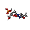  Type: DNA linking / Mass: 322.208 Da / Num. of mol.: 1 / Source method: obtained synthetically / Formula: C10H15N2O8P Type: DNA linking / Mass: 322.208 Da / Num. of mol.: 1 / Source method: obtained synthetically / Formula: C10H15N2O8PHas ligand of interest | N | Has protein modification | Y | |
|---|
-Experimental details
-Experiment
| Experiment | Method: ELECTRON MICROSCOPY |
|---|---|
| EM experiment | Aggregation state: PARTICLE / 3D reconstruction method: single particle reconstruction |
- Sample preparation
| Component |
| ||||||||||||||||||||||||
|---|---|---|---|---|---|---|---|---|---|---|---|---|---|---|---|---|---|---|---|---|---|---|---|---|---|
| Molecular weight | Experimental value: NO | ||||||||||||||||||||||||
| Source (natural) |
| ||||||||||||||||||||||||
| Source (recombinant) | Organism: Homo sapiens (human) | ||||||||||||||||||||||||
| Buffer solution | pH: 5.5 | ||||||||||||||||||||||||
| Specimen | Embedding applied: NO / Shadowing applied: NO / Staining applied: NO / Vitrification applied: YES | ||||||||||||||||||||||||
| Specimen support | Grid material: COPPER / Grid type: Quantifoil R1.2/1.3 | ||||||||||||||||||||||||
| Vitrification | Instrument: FEI VITROBOT MARK IV / Cryogen name: ETHANE / Humidity: 100 % / Chamber temperature: 279 K |
- Electron microscopy imaging
| Experimental equipment |  Model: Titan Krios / Image courtesy: FEI Company |
|---|---|
| Microscopy | Model: TFS KRIOS |
| Electron gun | Electron source:  FIELD EMISSION GUN / Accelerating voltage: 300 kV / Illumination mode: OTHER FIELD EMISSION GUN / Accelerating voltage: 300 kV / Illumination mode: OTHER |
| Electron lens | Mode: BRIGHT FIELD / Nominal magnification: 105000 X / Nominal defocus max: 2000 nm / Nominal defocus min: 1000 nm |
| Image recording | Electron dose: 48 e/Å2 / Film or detector model: GATAN K3 BIOQUANTUM (6k x 4k) |
- Processing
| EM software | Name: cryoSPARC / Category: 3D reconstruction |
|---|---|
| CTF correction | Type: PHASE FLIPPING AND AMPLITUDE CORRECTION |
| Symmetry | Point symmetry: C1 (asymmetric) |
| 3D reconstruction | Resolution: 3.06 Å / Resolution method: FSC 0.143 CUT-OFF / Num. of particles: 65742 / Symmetry type: POINT |
| Refinement | Cross valid method: NONE |
