 Movie
Movie Controller


+ Open data

- Basic information
| Entry |  | ||||||||||||
|---|---|---|---|---|---|---|---|---|---|---|---|---|---|
| Title | Cryo-EM structure of human PLD3 apo form | ||||||||||||
 Map data Map data | |||||||||||||
 Sample Sample |
| ||||||||||||
 Keywords Keywords | Exonuclease / IMMUNE SYSTEM | ||||||||||||
| Function / homology |  Function and homology information Function and homology informationspleen exonuclease / Synthesis of PG / single-stranded DNA 5'-3' DNA exonuclease activity / myotube differentiation / Hydrolases; Acting on ester bonds; Phosphoric-diester hydrolases / D-type glycerophospholipase activity / regulation of cytokine production involved in inflammatory response / Role of phospholipids in phagocytosis / immune system process / endomembrane system ...spleen exonuclease / Synthesis of PG / single-stranded DNA 5'-3' DNA exonuclease activity / myotube differentiation / Hydrolases; Acting on ester bonds; Phosphoric-diester hydrolases / D-type glycerophospholipase activity / regulation of cytokine production involved in inflammatory response / Role of phospholipids in phagocytosis / immune system process / endomembrane system / lysosomal lumen / lipid metabolic process / late endosome membrane / early endosome membrane / inflammatory response / Golgi membrane / lysosomal membrane / endoplasmic reticulum membrane / extracellular exosome Similarity search - Function | ||||||||||||
| Biological species |  Homo sapiens (human) Homo sapiens (human) | ||||||||||||
| Method | single particle reconstruction / cryo EM / Resolution: 2.9 Å | ||||||||||||
 Authors Authors | Hirano Y / Ezaki W / Ohto U / Shimizu T | ||||||||||||
| Funding support |  Japan, 3 items Japan, 3 items
| ||||||||||||
 Citation Citation | Journal: Nat Commun / Year: 2025 Title: Mechanistic insights into single-stranded DNA degradation by lysosomal exonucleases PLD3 and PLD4 from structural snapshots. Authors: Yoshinori Hirano / Wakiko Ezaki / Ryota Sato / Umeharu Ohto / Kensuke Miyake / Toshiyuki Shimizu / Abstract: Lysosomal exonuclease phospholipase D (PLD) family PLD3 and PLD4 degrade single-stranded RNA or DNA and regulate TLR7 or TLR9 responses. Polymorphisms of these enzymes are associated with human ...Lysosomal exonuclease phospholipase D (PLD) family PLD3 and PLD4 degrade single-stranded RNA or DNA and regulate TLR7 or TLR9 responses. Polymorphisms of these enzymes are associated with human diseases: PLD4 is associated with inflammatory diseases, and PLD3 is associated with neurodegenerative diseases. Here, we determine the structures of substrate-bound PLD3 and PLD4 by cryo-electron microscopy. Our structures reveal that PLD3 rebuilds a substrate-binding pocket, depending on the substrate, mainly via motion of the Phe335-containing loop. Furthermore, we captured the structure in a metastable state that appears during substrate rearrangement following product release. Together, our findings identify the residues that underlie the distinct activities of PLD3 and PLD4. This study provides a mechanistic basis for the exonuclease activity of PLD3 and PLD4 in single-stranded DNA degradation. | ||||||||||||
| History |
|
- Structure visualization
| Supplemental images |
|---|

- Downloads & links
-EMDB archive
| Map data | emd_64921.map.gz | 62.2 MB | EMDB map data format | |
|---|---|---|---|---|
| Header (meta data) | emd-64921-v30.xmlemd-64921.xml | 18.4 KB 18.4 KB | Display Display | EMDB header |
| Images |  emd_64921.png emd_64921.png | 63.6 KB | ||
| Filedesc metadata | emd-64921.cif.gz | 6.3 KB | ||
| Others | emd_64921_half_map_1.map.gzemd_64921_half_map_2.map.gz | 115.9 MB 115.9 MB | ||
| Archive directory |  http://ftp.pdbj.org/pub/emdb/structures/EMD-64921ftp://ftp.pdbj.org/pub/emdb/structures/EMD-64921 http://ftp.pdbj.org/pub/emdb/structures/EMD-64921ftp://ftp.pdbj.org/pub/emdb/structures/EMD-64921 | HTTPS FTP |
-Related structure data
| Related structure data |  9vbgMC  9vbhC  9vbiC  9vbjC  9vbkC M: atomic model generated by this map C: citing same article ( |
|---|---|
| Similar structure data |


-Links
| EMDB pages | EMDB (EBI/PDBe) / EMDataResource |
|---|---|
| Related items in Molecule of the Month |


-Map
| File | Download / File: emd_64921.map.gz / Format: CCP4 / Size: 125 MB / Type: IMAGE STORED AS FLOATING POINT NUMBER (4 BYTES) | ||||||||||||||||||||||||||||||||||||
|---|---|---|---|---|---|---|---|---|---|---|---|---|---|---|---|---|---|---|---|---|---|---|---|---|---|---|---|---|---|---|---|---|---|---|---|---|---|
| Projections & slices | Image control
Images are generated by Spider. | ||||||||||||||||||||||||||||||||||||
| Voxel size | X=Y=Z: 0.83 Å | ||||||||||||||||||||||||||||||||||||
| Density |
| ||||||||||||||||||||||||||||||||||||
| Symmetry | Space group: 1 | ||||||||||||||||||||||||||||||||||||
| Details | EMDB XML:
|
 Z (Sec.)
 Y (Row.)
 X (Col.)


-Supplemental data
-Half map: #2
| File | emd_64921_half_map_1.map | ||||||||||||
|---|---|---|---|---|---|---|---|---|---|---|---|---|---|
| Projections & Slices |
| ||||||||||||
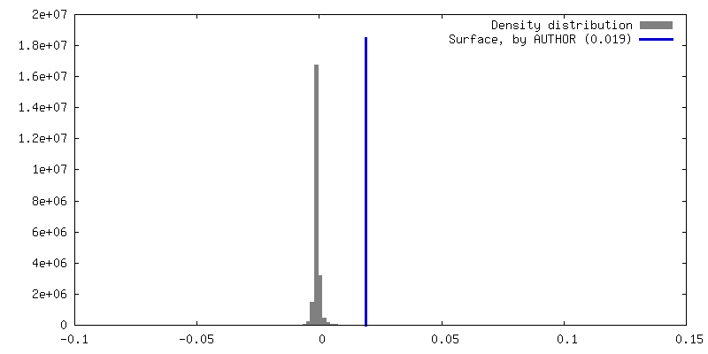
| Density Histograms |


-Half map: #1
| File | emd_64921_half_map_2.map | ||||||||||||
|---|---|---|---|---|---|---|---|---|---|---|---|---|---|
| Projections & Slices |
| ||||||||||||
| Density Histograms |


- Sample components
-Entire : Homodimer of PLD3 luminal domain
| Entire | Name: Homodimer of PLD3 luminal domain |
|---|---|
| Components |
|
-Supramolecule #1: Homodimer of PLD3 luminal domain
| Supramolecule | Name: Homodimer of PLD3 luminal domain / type: complex / ID: 1 / Parent: 0 / Macromolecule list: #1 |
|---|---|
| Source (natural) | Organism: Homo sapiens (human) |
-Macromolecule #1: 5'-3' exonuclease PLD3
| Macromolecule | Name: 5'-3' exonuclease PLD3 / type: protein_or_peptide / ID: 1 Details: C-terminal 6 residues (LEVLFQ) are derived from the expression tag. Number of copies: 2 / Enantiomer: LEVO / EC number: spleen exonuclease |
|---|---|
| Source (natural) | Organism: Homo sapiens (human) |
| Molecular weight | Theoretical: 48.601664 KDa |
| Recombinant expression | Organism: Homo sapiens (human) |
| Sequence | String: EYGDLHLFGP NQRPAPCYDP CEAVLVESIP EGLDFPNAST GNPSTSQAWL GLLAGAHSSL DIASFYWTLT NNDTHTQEPS AQQGEEVLR QLQTLAPKGV NVRIAVSKPS GPQPQADLQA LLQSGAQVRM VDMQKLTHGV LHTKFWVVDQ THFYLGSANM D WRSLTQVK ...String: EYGDLHLFGP NQRPAPCYDP CEAVLVESIP EGLDFPNAST GNPSTSQAWL GLLAGAHSSL DIASFYWTLT NNDTHTQEPS AQQGEEVLR QLQTLAPKGV NVRIAVSKPS GPQPQADLQA LLQSGAQVRM VDMQKLTHGV LHTKFWVVDQ THFYLGSANM D WRSLTQVK ELGVVMYNCS CLARDLTKIF EAYWFLGQAG SSIPSTWPRF YDTRYNQETP MEICLNGTPA LAYLASAPPP LC PSGRTPD LKALLNVVDN ARSFIYVAVM NYLPTLEFSH PHRFWPAIDD GLRRATYERG VKVRLLISCW GHSEPSMRAF LLS LAALRD NHTHSDIQVK LFVVPADEAQ ARIPYARVNH NKYMVTERAT YIGTSNWSGN YFTETAGTSL LVTQNGRGGL RSQL EAIFL RDWDSPYSHD LDTSADSVGN ACRLLLEVLF Q UniProtKB: 5'-3' exonuclease PLD3 |
-Macromolecule #5: 2-acetamido-2-deoxy-beta-D-glucopyranose
| Macromolecule | Name: 2-acetamido-2-deoxy-beta-D-glucopyranose / type: ligand / ID: 5 / Number of copies: 4 / Formula: NAG |
|---|---|
| Molecular weight | Theoretical: 221.208 Da |
| Chemical component information |  ChemComp-NAG: |
-Experimental details
-Structure determination
| Method | cryo EM |
|---|---|
 Processing Processing | single particle reconstruction |
| Aggregation state | particle |
-Sample preparation
| Buffer | pH: 7 |
|---|---|
| Grid | Model: Quantifoil R1.2/1.3 / Material: COPPER / Pretreatment - Type: GLOW DISCHARGE |
| Vitrification | Cryogen name: ETHANE / Chamber humidity: 100 % / Chamber temperature: 279 K / Instrument: FEI VITROBOT MARK IV |
- Electron microscopy
| Microscope | TFS KRIOS |
|---|---|
| Image recording | Film or detector model: GATAN K3 BIOCONTINUUM (6k x 4k) / Average electron dose: 61.0 e/Å2 |
| Electron beam | Acceleration voltage: 300 kV / Electron source:  FIELD EMISSION GUN FIELD EMISSION GUN |
| Electron optics | Illumination mode: OTHER / Imaging mode: BRIGHT FIELD / Nominal defocus max: 2.0 µm / Nominal defocus min: 0.8 µm / Nominal magnification: 105000 |
| Experimental equipment |  Model: Titan Krios / Image courtesy: FEI Company |
