 Movie
Movie Controller


+ Open data

- Basic information
| Entry | Database: PDB / ID: 9tlg | |||||||||||||||||||||||||||||||||
|---|---|---|---|---|---|---|---|---|---|---|---|---|---|---|---|---|---|---|---|---|---|---|---|---|---|---|---|---|---|---|---|---|---|---|
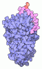
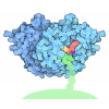
| Title | Structure of human prothrombinase with meizothrombin | |||||||||||||||||||||||||||||||||
 Components Components |
| |||||||||||||||||||||||||||||||||
 Keywords Keywords | BLOOD CLOTTING / prothrombinase / factor Va / factor Xa / meizothrombin | |||||||||||||||||||||||||||||||||
| Function / homology |  Function and homology information Function and homology informationDefective cleavage of FV variant at a.a.534 / Defective cleavage of FV variant at R334 / response to vitamin K / coagulation factor Xa / platelet alpha granule / Cargo concentration in the ER / COPII-coated ER to Golgi transport vesicle / Defective factor IX causes thrombophilia / Defective cofactor function of FVIIIa variant / Defective F9 variant does not activate FX ...Defective cleavage of FV variant at a.a.534 / Defective cleavage of FV variant at R334 / response to vitamin K / coagulation factor Xa / platelet alpha granule / Cargo concentration in the ER / COPII-coated ER to Golgi transport vesicle / Defective factor IX causes thrombophilia / Defective cofactor function of FVIIIa variant / Defective F9 variant does not activate FX / COPII-mediated vesicle transport / blood circulation / negative regulation of astrocyte differentiation / thrombospondin receptor activity / thrombin / thrombin-activated receptor signaling pathway / Defective factor XII causes hereditary angioedema / regulation of blood coagulation / neutrophil-mediated killing of gram-negative bacterium / positive regulation of phospholipase C-activating G protein-coupled receptor signaling pathway / Platelet Aggregation (Plug Formation) / Defective F8 cleavage by thrombin / positive regulation of collagen biosynthetic process / negative regulation of platelet activation / negative regulation of blood coagulation / Fibrin formation / negative regulation of fibrinolysis / positive regulation of blood coagulation / negative regulation of proteolysis / Transport of gamma-carboxylated protein precursors from the endoplasmic reticulum to the Golgi apparatus / fibrinolysis / Gamma-carboxylation of protein precursors / Removal of aminoterminal propeptides from gamma-carboxylated proteins / regulation of cytosolic calcium ion concentration / Initiation of coagulation cascade / Amplification and propagation of coagulation cascade / negative regulation of cytokine production involved in inflammatory response / endoplasmic reticulum-Golgi intermediate compartment membrane / positive regulation of TOR signaling / platelet alpha granule lumen / Regulation of Complement cascade / positive regulation of receptor signaling pathway via JAK-STAT / positive regulation of release of sequestered calcium ion into cytosol / growth factor activity / Cell surface interactions at the vascular wall / Regulation of clotting cascade / Peptide ligand-binding receptors / Post-translational protein phosphorylation / response to wounding / lipopolysaccharide binding / platelet activation / blood coagulation / positive regulation of protein localization to nucleus / phospholipid binding / Golgi lumen / Regulation of Insulin-like Growth Factor (IGF) transport and uptake by Insulin-like Growth Factor Binding Proteins (IGFBPs) / positive regulation of reactive oxygen species metabolic process / heparin binding / Platelet degranulation / extracellular vesicle / Thrombin signalling through proteinase activated receptors (PARs) / Dengue Virus-Host Interactions / antimicrobial humoral immune response mediated by antimicrobial peptide / blood microparticle / extracellular matrix / G alpha (q) signalling events / cell surface receptor signaling pathway / positive regulation of cell migration / receptor ligand activity / endoplasmic reticulum lumen / copper ion binding / serine-type endopeptidase activity / external side of plasma membrane / signaling receptor binding / calcium ion binding / proteolysis / extracellular exosome / extracellular region / membrane / plasma membrane Similarity search - Function | |||||||||||||||||||||||||||||||||
| Biological species |  Homo sapiens (human) Homo sapiens (human) | |||||||||||||||||||||||||||||||||
| Method | ELECTRON MICROSCOPY / single particle reconstruction / cryo EM / Resolution: 3.1 Å | |||||||||||||||||||||||||||||||||
 Authors Authors | Faille, A. / Ustok, F.I. / Warren, A. / Huntingon, J.A. | |||||||||||||||||||||||||||||||||
| Funding support |  United Kingdom, 3items United Kingdom, 3items
| |||||||||||||||||||||||||||||||||
 Citation Citation | Journal: EMBO J / Year: 2026 Title: Prothrombinase processivity is conferred by substrate allostery. Authors: Fatma Işık Üstok / Alexandre Faille / Alan J Warren / James A Huntington / Abstract: The prothrombinase complex, comprised of factor (f) Xa and fVa, converts prothrombin to thrombin through sequential cleavage at two sites in a rapid and processive manner. The molecular basis of ...The prothrombinase complex, comprised of factor (f) Xa and fVa, converts prothrombin to thrombin through sequential cleavage at two sites in a rapid and processive manner. The molecular basis of prothrombin processing is an enzymatical mystery that to solve requires structural insight into how the substrate and intermediate bind to prothrombinase. Here we present two 3.1 Å cryo-EM structures of prothrombinase bound to prothrombin and to meizothrombin. The prothrombin complex revealed a surprising interaction between the end of the heavy chain of fVa with exosite I of prothrombin, accounting for 70% of the contact interface. Triggering of the zymogen-to-protease conformational change following cleavage at Arg320 alters all domain-domain and fVa interactions observed for prothrombin, and results in a large-scale rearrangement of meizothrombin that presents the second cleavage site (Arg271) for processing. Together, these structures reveal a remarkable enzymatic mechanism that requires the active participation of the substrate itself, and introduces a new paradigm of 'substrate allostery'. | |||||||||||||||||||||||||||||||||
| History |
|
- Structure visualization
| Structure viewer | Molecule: MolmilJmol/JSmol |
|---|
- Downloads & links
-Download
| PDBx/mmCIF format | 9tlg.cif.gz | 531.9 KB | Display | PDBx/mmCIF format |
|---|---|---|---|---|
| PDB format | pdb9tlg.ent.gz | 339.9 KB | Display | PDB format |
| PDBx/mmJSON format | 9tlg.json.gz | Tree view | PDBx/mmJSON format | |
| Others |  Other downloads Other downloads |
-Validation report
| Arichive directory | https://data.pdbj.org/pub/pdb/validation_reports/tl/9tlgftp://data.pdbj.org/pub/pdb/validation_reports/tl/9tlg | HTTPS FTP |
|---|
-Related structure data
| Related structure data |  56054MC  9t00C  9tleC M: map data used to model this data C: citing same article ( |
|---|---|
| Similar structure data |


-Links
 PDBj
PDBj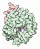






- Assembly
| Deposited unit | 
|
|---|---|
| 1 |
|
-Components
-Coagulation factor V ... , 2 types, 2 molecules AB
| #1: Protein | Mass: 81514.578 Da / Num. of mol.: 1 Source method: isolated from a genetically manipulated source Source: (gene. exp.) Homo sapiens (human) / Gene: F5 / Production host:   Escherichia coli (E. coli) / References: UniProt: P12259 Escherichia coli (E. coli) / References: UniProt: P12259 |
|---|---|
| #2: Protein | Mass: 75283.008 Da / Num. of mol.: 1 Source method: isolated from a genetically manipulated source Source: (gene. exp.) Homo sapiens (human) / Gene: F5 / Production host: Escherichia coli (E. coli) / References: UniProt: P12259 |
-Protein , 4 types, 4 molecules HLPQ
| #3: Protein | Mass: 28773.916 Da / Num. of mol.: 1 Mutation: E129N, S130E, T132K, K134D, N166H, K169M, S173D, I175R, S195A, A233R, D239K Source method: isolated from a genetically manipulated source Source: (gene. exp.) Homo sapiens (human) / Gene: F10 / Production host: Escherichia coli (E. coli) / References: UniProt: P00742 |
|---|---|
| #4: Protein | Mass: 12182.554 Da / Num. of mol.: 1 Mutation: T85M, S90R, L91A, D92F, H101K, E102R, E103V, N105S Source method: isolated from a genetically manipulated source Details: M17 construct with 17 mutations enhancing affinity towards factor V, first 85 residues truncated, catalytic mutation S195A Source: (gene. exp.) Homo sapiens (human) / Gene: F10 / Production host: Escherichia coli (E. coli) / References: UniProt: P00742, coagulation factor Xa |
| #5: Protein | Mass: 35623.961 Da / Num. of mol.: 1 Source method: isolated from a genetically manipulated source Source: (gene. exp.) Homo sapiens (human) / Gene: F2 / Cell line (production host): HEK293 / Production host: Homo sapiens (human) / References: UniProt: P00734, thrombin |
| #6: Protein | Mass: 29764.219 Da / Num. of mol.: 1 Source method: isolated from a genetically manipulated source Details: S195A mutation / Source: (gene. exp.) Homo sapiens (human) / Gene: F2 / Cell line (production host): HEK293 / Production host: Homo sapiens (human) / References: UniProt: P00734 |
-Sugars , 4 types, 5 molecules 
| #7: Polysaccharide | beta-D-mannopyranose-(1-4)-2-acetamido-2-deoxy-beta-D-glucopyranose-(1-4)-2-acetamido-2-deoxy-beta- ...beta-D-mannopyranose-(1-4)-2-acetamido-2-deoxy-beta-D-glucopyranose-(1-4)-2-acetamido-2-deoxy-beta-D-glucopyranose Source method: isolated from a genetically manipulated source | ||||
|---|---|---|---|---|---|
| #8: Polysaccharide | Source method: isolated from a genetically manipulated source #9: Polysaccharide | alpha-D-mannopyranose-(1-6)-alpha-D-mannopyranose-(1-3)-[alpha-D-mannopyranose-(1-6)-alpha-D- ...alpha-D-mannopyranose-(1-6)-alpha-D-mannopyranose-(1-3)-[alpha-D-mannopyranose-(1-6)-alpha-D-mannopyranose-(1-6)]beta-D-mannopyranose-(1-4)-2-acetamido-2-deoxy-beta-D-glucopyranose-(1-4)-2-acetamido-2-deoxy-beta-D-glucopyranose | Type: oligosaccharide / Mass: 1235.105 Da / Num. of mol.: 1 Source method: isolated from a genetically manipulated source #10: Sugar | ChemComp-NAG / |  Type: D-saccharide, beta linking / Mass: 221.208 Da / Num. of mol.: 1 / Source method: obtained synthetically / Formula: C8H15NO6 Type: D-saccharide, beta linking / Mass: 221.208 Da / Num. of mol.: 1 / Source method: obtained synthetically / Formula: C8H15NO6 |
-Non-polymers , 2 types, 3 molecules 

| #11: Chemical | ChemComp-CA /  Mass: 40.078 Da / Num. of mol.: 1 / Source method: obtained synthetically / Formula: Ca Mass: 40.078 Da / Num. of mol.: 1 / Source method: obtained synthetically / Formula: Ca |
|---|---|
| #12: Chemical |  Mass: 22.990 Da / Num. of mol.: 2 / Source method: obtained synthetically / Formula: Na Mass: 22.990 Da / Num. of mol.: 2 / Source method: obtained synthetically / Formula: Na |
-Details
| Has ligand of interest | N |
|---|---|
| Has protein modification | Y |
-Experimental details
-Experiment
| Experiment | Method: ELECTRON MICROSCOPY |
|---|---|
| EM experiment | Aggregation state: PARTICLE / 3D reconstruction method: single particle reconstruction |
- Sample preparation
| Component | Name: Prothrombinase in complex with meizothrombin / Type: COMPLEX / Entity ID: #1-#6 / Source: RECOMBINANT |
|---|---|
| Source (natural) | Organism: Homo sapiens (human) |
| Source (recombinant) | Organism: Homo sapiens (human) / Strain: HEK293 |
| Buffer solution | pH: 7.4 |
| Specimen | Embedding applied: NO / Shadowing applied: NO / Staining applied: NO / Vitrification applied: YES |
| Vitrification | Cryogen name: ETHANE |
- Electron microscopy imaging
| Experimental equipment |  Model: Titan Krios / Image courtesy: FEI Company |
|---|---|
| Microscopy | Model: TFS KRIOS |
| Electron gun | Electron source:  FIELD EMISSION GUN / Accelerating voltage: 300 kV / Illumination mode: FLOOD BEAM FIELD EMISSION GUN / Accelerating voltage: 300 kV / Illumination mode: FLOOD BEAM |
| Electron lens | Mode: BRIGHT FIELD / Nominal defocus max: 1800 nm / Nominal defocus min: 600 nm |
| Image recording | Electron dose: 52.7 e/Å2 / Film or detector model: FEI FALCON IV (4k x 4k) |
- Processing
| EM software |
| ||||||||||||||||||||||||
|---|---|---|---|---|---|---|---|---|---|---|---|---|---|---|---|---|---|---|---|---|---|---|---|---|---|
| CTF correction | Type: PHASE FLIPPING AND AMPLITUDE CORRECTION | ||||||||||||||||||||||||
| 3D reconstruction | Resolution: 3.1 Å / Resolution method: FSC 0.143 CUT-OFF / Num. of particles: 25376 / Symmetry type: POINT | ||||||||||||||||||||||||
| Refinement | Cross valid method: NONE Stereochemistry target values: GeoStd + Monomer Library + CDL v1.2 | ||||||||||||||||||||||||
| Displacement parameters | Biso mean: 59.8 Å2 | ||||||||||||||||||||||||
| Refine LS restraints |
|
