 Movie
Movie Controller


+ Open data

- Basic information
| Entry | Database: PDB / ID: 9t65 | |||||||||||||||||||||||||||
|---|---|---|---|---|---|---|---|---|---|---|---|---|---|---|---|---|---|---|---|---|---|---|---|---|---|---|---|---|
| Title | Complex linking two cytoplasmic lattice filaments | |||||||||||||||||||||||||||
 Components Components |
| |||||||||||||||||||||||||||
 Keywords Keywords | UNKNOWN FUNCTION / Ubiquitination / Complex / Filament | |||||||||||||||||||||||||||
| Function / homology |  Function and homology information Function and homology informationregulation of translation by machinery localization / cytoplasm organization / ooplasm / Prolactin receptor signaling / Chromatin modifying enzymes / protein storage / structural constituent of cytoplasmic lattice / cytoplasmic lattice / SCF-beta-TrCP mediated degradation of Emi1 / Regulation of BACH1 activity ...regulation of translation by machinery localization / cytoplasm organization / ooplasm / Prolactin receptor signaling / Chromatin modifying enzymes / protein storage / structural constituent of cytoplasmic lattice / cytoplasmic lattice / SCF-beta-TrCP mediated degradation of Emi1 / Regulation of BACH1 activity / histone H3K18 ubiquitin ligase activity / histone H3 ubiquitin ligase activity / histone H3K14 ubiquitin ligase activity / histone H3K23 ubiquitin ligase activity / SCF(Skp2)-mediated degradation of p27/p21 / MAP3K8 (TPL2)-dependent MAPK1/3 activation / histone H3 reader activity / Regulation of RUNX2 expression and activity / Degradation of GLI1 by the proteasome / GSK3B-mediated proteasomal degradation of PD-L1(CD274) / Cyclin D associated events in G1 / FBXL7 down-regulates AURKA during mitotic entry and in early mitosis / cortical granule / Orc1 removal from chromatin / GSK3B and BTRC:CUL1-mediated-degradation of NFE2L2 / Dectin-1 mediated noncanonical NF-kB signaling / NIK-->noncanonical NF-kB signaling / Degradation of beta-catenin by the destruction complex / Activation of NF-kappaB in B cells / Iron uptake and transport / embryonic cleavage / chromosomal DNA methylation maintenance following DNA replication / intermediate filament cytoskeleton / CLEC7A (Dectin-1) signaling / FCERI mediated NF-kB activation / Interleukin-1 signaling / F-box domain binding / Downstream TCR signaling / hemi-methylated DNA-binding / GLI3 is processed to GLI3R by the proteasome / Regulation of PLK1 Activity at G2/M Transition / regulation of epithelial cell proliferation / Ubiquitin-Mediated Degradation of Phosphorylated Cdc25A / Neddylation / PcG protein complex / gap junction / maintenance of protein location in nucleus / positive regulation of epithelial cell apoptotic process / Cul7-RING ubiquitin ligase complex / methyl-CpG binding / Antigen processing: Ubiquitination & Proteasome degradation / ubiquitin ligase activator activity / epigenetic programming in the zygotic pronuclei / histone H3K9me2/3 reader activity / negative regulation of gene expression via chromosomal CpG island methylation / SCF ubiquitin ligase complex / SCF-dependent proteasomal ubiquitin-dependent protein catabolic process / positive regulation of protein metabolic process / ubiquitin ligase complex scaffold activity / mitotic spindle assembly / protein monoubiquitination / cullin family protein binding / cis-regulatory region sequence-specific DNA binding / protein autoubiquitination / ubiquitin-like ligase-substrate adaptor activity / cytoskeleton organization / protein localization to chromatin / heterochromatin / protein K48-linked ubiquitination / in utero embryonic development / replication fork / molecular function activator activity / euchromatin / tubulin binding / RING-type E3 ubiquitin transferase / beta-catenin binding / intracellular protein localization / nuclear matrix / protein polyubiquitination / ubiquitin-protein transferase activity / ubiquitin protein ligase activity / regulation of inflammatory response / heterochromatin formation / spermatogenesis / ubiquitin-dependent protein catabolic process / histone binding / nucleic acid binding / proteasome-mediated ubiquitin-dependent protein catabolic process / cell differentiation / protein ubiquitination / chromatin remodeling / protein domain specific binding / DNA repair / apoptotic process / centrosome / calcium ion binding / chromatin / negative regulation of transcription by RNA polymerase II / positive regulation of transcription by RNA polymerase II / DNA-templated transcription Similarity search - Function | |||||||||||||||||||||||||||
| Biological species |   Mus musculus (house mouse) Mus musculus (house mouse) | |||||||||||||||||||||||||||
| Method | ELECTRON MICROSCOPY / subtomogram averaging / cryo EM / Resolution: 5.6 Å | |||||||||||||||||||||||||||
 Authors Authors | Singh, K. / Harasimov, K. / Carter, A.P. | |||||||||||||||||||||||||||
| Funding support |  United Kingdom, European Union, 3items United Kingdom, European Union, 3items
| |||||||||||||||||||||||||||
 Citation Citation | Journal: EMBO J / Year: 2026 Title: In-situ cryo-ET of mouse embryos reveals cytoplasmic lattices contain ubiquitin-charged E2-E3 ligase assemblies. Authors: Kashish Singh / Katarina Harasimov / Kathy K Niakan / Andrew P Carter / Abstract: Cytoplasmic lattices (CPLs) are filamentous assemblies essential for mammalian embryonic development. They are known to regulate organelle organization, spindle assembly, and protein homeostasis, but ...Cytoplasmic lattices (CPLs) are filamentous assemblies essential for mammalian embryonic development. They are known to regulate organelle organization, spindle assembly, and protein homeostasis, but their molecular functions remain unclear. Here, we develop a strategy combining cryo-focused ion beam milling and cryo-electron tomography to resolve macromolecular complexes directly in mammalian embryos. Using this approach, we determine the in situ structure of cytoplasmic lattices within 6/8-cell mouse embryos at ~4.7 Å resolution. CPL filaments are built from multiple copies of at least fourteen proteins arranged into a ~4.5 MDa repeating unit. The repeat contains a central cavity that is open at the back and lined with multiple FBXW-SKP1 complexes and three modules, each containing the E2 ubiquitin-conjugating enzyme UBE2D and the E3 ligase UHRF1. We resolve two CPL states: one is consistent with a ubiquitin-charged UBE2D, where ubiquitin is held in an open, inactive conformation by binding the scaffold protein PADI6; the second lacks discernible ubiquitin density and shows structural changes compatible with ubiquitin becoming available for transfer. Our findings support a model in which CPLs function as large ubiquitin ligase assemblies during early embryonic development. | |||||||||||||||||||||||||||
| History |
|
- Structure visualization
| Structure viewer | Molecule: MolmilJmol/JSmol |
|---|
- Downloads & links
-Download
| PDBx/mmCIF format | 9t65.cif.gz | 966.1 KB | Display | PDBx/mmCIF format |
|---|---|---|---|---|
| PDB format | pdb9t65.ent.gz | 696.7 KB | Display | PDB format |
| PDBx/mmJSON format | 9t65.json.gz | Tree view | PDBx/mmJSON format | |
| Others |  Other downloads Other downloads |
-Validation report
| Arichive directory | https://data.pdbj.org/pub/pdb/validation_reports/t6/9t65ftp://data.pdbj.org/pub/pdb/validation_reports/t6/9t65 | HTTPS FTP |
|---|
-Related structure data
| Related structure data |  55605MC  9t63C  9t64C  9t66C  9t67C  9t68C M: map data used to model this data C: citing same article ( |
|---|---|
| Similar structure data |


-Links
 PDBj
PDBj
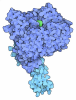


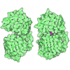

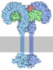
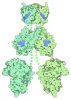

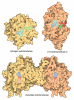
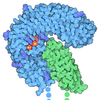
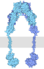
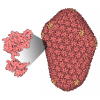


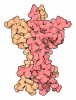
- Assembly
| Deposited unit | 
|
|---|---|
| 1 |
|
-Components
| #1: Protein | Mass: 76854.109 Da / Num. of mol.: 8 / Source method: isolated from a natural source / Source: (natural) Mus musculus (house mouse) / References: UniProt: Q8K3V4#2: Protein | | Mass: 18693.992 Da / Num. of mol.: 1 / Source method: isolated from a natural source / Source: (natural) Mus musculus (house mouse) / References: UniProt: Q9WTX5#3: Protein | | Mass: 113527.188 Da / Num. of mol.: 1 / Source method: isolated from a natural source / Source: (natural) Mus musculus (house mouse) / References: UniProt: Q6B966#4: Protein | | Mass: 88436.805 Da / Num. of mol.: 1 / Source method: isolated from a natural source / Source: (natural) Mus musculus (house mouse)References: UniProt: Q8VDF2, RING-type E3 ubiquitin transferase #5: Protein | | Mass: 39676.895 Da / Num. of mol.: 1 / Source method: isolated from a natural source / Source: (natural) Mus musculus (house mouse)Has protein modification | N | |
|---|
-Experimental details
-Experiment
| Experiment | Method: ELECTRON MICROSCOPY |
|---|---|
| EM experiment | Aggregation state: FILAMENT / 3D reconstruction method: subtomogram averaging |
- Sample preparation
| Component | Name: Multi-subunit complex containing PADI6, SKP1, a F-box/WD repeat-containing protein, NLRP14 and UHRF1 Type: COMPLEX / Entity ID: all / Source: NATURAL |
|---|---|
| Source (natural) | Organism: Mus musculus (house mouse) |
| Buffer solution | pH: 7.5 |
| Specimen | Embedding applied: NO / Shadowing applied: NO / Staining applied: NO / Vitrification applied: YES |
| Vitrification | Cryogen name: ETHANE |
- Electron microscopy imaging
| Experimental equipment |  Model: Titan Krios / Image courtesy: FEI Company |
|---|---|
| Microscopy | Model: TFS KRIOS |
| Electron gun | Electron source:  FIELD EMISSION GUN / Accelerating voltage: 300 kV / Illumination mode: FLOOD BEAM FIELD EMISSION GUN / Accelerating voltage: 300 kV / Illumination mode: FLOOD BEAM |
| Electron lens | Mode: BRIGHT FIELD / Nominal defocus max: 4000 nm / Nominal defocus min: 1000 nm |
| Image recording | Electron dose: 3.5 e/Å2 / Avg electron dose per subtomogram: 115 e/Å2 / Film or detector model: FEI FALCON IV (4k x 4k) |
- Processing
| EM software |
| ||||||||||||
|---|---|---|---|---|---|---|---|---|---|---|---|---|---|
| CTF correction | Type: PHASE FLIPPING AND AMPLITUDE CORRECTION | ||||||||||||
| Symmetry | Point symmetry: C1 (asymmetric) | ||||||||||||
| 3D reconstruction | Resolution: 5.6 Å / Resolution method: FSC 0.143 CUT-OFF / Num. of particles: 86696 / Symmetry type: POINT | ||||||||||||
| EM volume selection | Num. of tomograms: 1153 / Num. of volumes extracted: 268774 |
