 Movie
Movie Controller

[English] 日本語
 Yorodumi
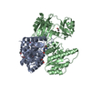
- PDB-9swj: Human ADP-forming succinyl-CoA ligase complex SUCLG1-SUCLA2 bound... -
+ Open data

- Basic information
| Entry | Database: PDB / ID: 9swj | ||||||||||||||||||||||||
|---|---|---|---|---|---|---|---|---|---|---|---|---|---|---|---|---|---|---|---|---|---|---|---|---|---|
| Title | Human ADP-forming succinyl-CoA ligase complex SUCLG1-SUCLA2 bound to coenzyme A | ||||||||||||||||||||||||
 Components Components |
| ||||||||||||||||||||||||
 Keywords Keywords | TRANSFERASE / Succinate / Complex / ATP / ADP / Coenzyme A | ||||||||||||||||||||||||
| Function / homology |  Function and homology information Function and homology informationsuccinyl-CoA pathway / succinate-CoA ligase activity / malate-CoA ligase / succinate-CoA ligase complex (GDP-forming) / malate-CoA ligase activity / succinate-CoA ligase (GDP-forming) / toxic metabolite repair / succinate-CoA ligase (GDP-forming) activity / succinate-CoA ligase complex (ADP-forming) / succinate-CoA ligase (ADP-forming) ...succinyl-CoA pathway / succinate-CoA ligase activity / malate-CoA ligase / succinate-CoA ligase complex (GDP-forming) / malate-CoA ligase activity / succinate-CoA ligase (GDP-forming) / toxic metabolite repair / succinate-CoA ligase (GDP-forming) activity / succinate-CoA ligase complex (ADP-forming) / succinate-CoA ligase (ADP-forming) / succinate-CoA ligase complex / succinate-CoA ligase (ADP-forming) activity / Ligases; Forming carbon-sulfur bonds; Acid-thiol ligases / succinyl-CoA catabolic process / succinyl-CoA metabolic process / succinate metabolic process / Citric acid cycle (TCA cycle) / malate metabolic process / catalytic complex / tricarboxylic acid cycle / mitochondrial matrix / nucleotide binding / magnesium ion binding / mitochondrion / RNA binding / extracellular exosome / ATP binding Similarity search - Function | ||||||||||||||||||||||||
| Biological species |  Homo sapiens (human) Homo sapiens (human) | ||||||||||||||||||||||||
| Method | ELECTRON MICROSCOPY / single particle reconstruction / cryo EM / Resolution: 3.7 Å | ||||||||||||||||||||||||
 Authors Authors | Bailey, H.J. / McCorvie, T.J. / Shrestha, L. / Rembeza, E. / Strain-Damerell, C. / Burgess-Brown, N. / Yue, W.W. | ||||||||||||||||||||||||
| Funding support |  United Kingdom, 1items United Kingdom, 1items
| ||||||||||||||||||||||||
 Citation Citation | Journal: To Be Published Title: Human ADP-forming succinyl-CoA ligase complex SUCLG1-SUCLA2 bound to coenzyme A Authors: Bailey, H.J. / McCorvie, T.J. / Yue, W.W. | ||||||||||||||||||||||||
| History |
|
- Structure visualization
| Structure viewer | Molecule: MolmilJmol/JSmol |
|---|
- Downloads & links
-Download
| PDBx/mmCIF format | 9swj.cif.gz | 484.3 KB | Display | PDBx/mmCIF format |
|---|---|---|---|---|
| PDB format | pdb9swj.ent.gz | 402 KB | Display | PDB format |
| PDBx/mmJSON format | 9swj.json.gz | Tree view | PDBx/mmJSON format | |
| Others |  Other downloads Other downloads |
-Validation report
| Arichive directory | https://data.pdbj.org/pub/pdb/validation_reports/sw/9swjftp://data.pdbj.org/pub/pdb/validation_reports/sw/9swj | HTTPS FTP |
|---|
-Related structure data
| Related structure data |  55309MC M: map data used to model this data C: citing same article ( |
|---|---|
| Similar structure data |
-Links
 PDBj
PDBj


- Assembly
| Deposited unit | 
|
|---|---|
| 1 |
|
-Components
| #1: Protein | Mass: 32161.875 Da / Num. of mol.: 4 Source method: isolated from a genetically manipulated source Source: (gene. exp.) Homo sapiens (human) / Gene: SUCLG1 / Production host:  References: UniProt: P53597, succinate-CoA ligase (GDP-forming), succinate-CoA ligase (ADP-forming) #2: Protein | Mass: 47757.012 Da / Num. of mol.: 4 Source method: isolated from a genetically manipulated source Source: (gene. exp.) Homo sapiens (human) / Gene: SUCLA2 / Production host: References: UniProt: Q9P2R7, succinate-CoA ligase (ADP-forming) #3: Chemical | ChemComp-COA /   Mass: 767.534 Da / Num. of mol.: 4 / Source method: obtained synthetically / Formula: C21H36N7O16P3S / Feature type: SUBJECT OF INVESTIGATION Mass: 767.534 Da / Num. of mol.: 4 / Source method: obtained synthetically / Formula: C21H36N7O16P3S / Feature type: SUBJECT OF INVESTIGATIONHas ligand of interest | Y | Has protein modification | N | |
|---|
-Experimental details
-Experiment
| Experiment | Method: ELECTRON MICROSCOPY |
|---|---|
| EM experiment | Aggregation state: PARTICLE / 3D reconstruction method: single particle reconstruction |
- Sample preparation
| Component | Name: Tetramer of heterodimers of SUCLG1-SUCLA2 bound to coenzyme A Type: COMPLEX / Entity ID: #1-#2 / Source: RECOMBINANT | ||||||||||||
|---|---|---|---|---|---|---|---|---|---|---|---|---|---|
| Molecular weight | Value: 0.313 MDa / Experimental value: NO | ||||||||||||
| Source (natural) | Organism: Homo sapiens (human) | ||||||||||||
| Source (recombinant) | Organism: | ||||||||||||
| Buffer solution | pH: 7.5 | ||||||||||||
| Buffer component |
| ||||||||||||
| Specimen | Conc.: 1 mg/ml / Embedding applied: NO / Shadowing applied: NO / Staining applied: NO / Vitrification applied: YES | ||||||||||||
| Specimen support | Grid material: GOLD / Grid mesh size: 200 divisions/in. / Grid type: Quantifoil R1.2/1.3 | ||||||||||||
| Vitrification | Instrument: FEI VITROBOT MARK III / Cryogen name: ETHANE / Humidity: 95 % / Chamber temperature: 277.15 K |
- Electron microscopy imaging
| Experimental equipment |  Model: Titan Krios / Image courtesy: FEI Company |
|---|---|
| Microscopy | Model: TFS KRIOS |
| Electron gun | Electron source:  FIELD EMISSION GUN / Accelerating voltage: 300 kV / Illumination mode: FLOOD BEAM FIELD EMISSION GUN / Accelerating voltage: 300 kV / Illumination mode: FLOOD BEAM |
| Electron lens | Mode: BRIGHT FIELD / Nominal magnification: 165000 X / Nominal defocus max: 2200 nm / Nominal defocus min: 1400 nm |
| Specimen holder | Cryogen: NITROGEN / Specimen holder model: FEI TITAN KRIOS AUTOGRID HOLDER |
| Image recording | Average exposure time: 4 sec. / Electron dose: 52.56 e/Å2 / Detector mode: COUNTING / Film or detector model: GATAN K2 QUANTUM (4k x 4k) / Num. of grids imaged: 1 / Num. of real images: 4100 |
| Image scans | Movie frames/image: 50 |
- Processing
| EM software |
| |||||||||||||||||||||||||||
|---|---|---|---|---|---|---|---|---|---|---|---|---|---|---|---|---|---|---|---|---|---|---|---|---|---|---|---|---|
| CTF correction | Type: PHASE FLIPPING AND AMPLITUDE CORRECTION | |||||||||||||||||||||||||||
| Particle selection | Num. of particles selected: 320000 | |||||||||||||||||||||||||||
| Symmetry | Point symmetry: D2 (2x2 fold dihedral) | |||||||||||||||||||||||||||
| 3D reconstruction | Resolution: 3.7 Å / Resolution method: FSC 0.143 CUT-OFF / Num. of particles: 44786 / Symmetry type: POINT | |||||||||||||||||||||||||||
| Atomic model building | Protocol: FLEXIBLE FIT / Space: REAL Details: Initially flexible fitting used the Namdinator server | |||||||||||||||||||||||||||
| Atomic model building | PDB-ID: 6G4Q Accession code: 6G4Q / Source name: PDB / Type: experimental model | |||||||||||||||||||||||||||
| Refinement | Highest resolution: 3.7 Å / Cross valid method: NONE Stereochemistry target values: REAL-SPACE (WEIGHTED MAP SUM AT ATOM CENTERS) |
