[English] 日本語
 Yorodumi
- EMDB-55309: Human ADP-forming succinyl-CoA ligase complex SUCLG1-SUCLA2 bound... -
+ Open data
- Basic information
| Entry |  | |||||||||
|---|---|---|---|---|---|---|---|---|---|---|
| Title | Human ADP-forming succinyl-CoA ligase complex SUCLG1-SUCLA2 bound to coenzyme A | |||||||||
 Map data Map data | ||||||||||
 Sample Sample |
| |||||||||
 Keywords Keywords | Succinate / Complex / ATP / ADP / Coenzyme A / TRANSFERASE | |||||||||
| Function / homology |  Function and homology information Function and homology informationsuccinyl-CoA pathway / succinate-CoA ligase complex (GDP-forming) / succinate-CoA ligase (GDP-forming) / succinate-CoA ligase (GDP-forming) activity / succinate-CoA ligase complex (ADP-forming) / succinate-CoA ligase (ADP-forming) / succinate-CoA ligase complex / succinate-CoA ligase (ADP-forming) activity / succinyl-CoA catabolic process / succinyl-CoA metabolic process ...succinyl-CoA pathway / succinate-CoA ligase complex (GDP-forming) / succinate-CoA ligase (GDP-forming) / succinate-CoA ligase (GDP-forming) activity / succinate-CoA ligase complex (ADP-forming) / succinate-CoA ligase (ADP-forming) / succinate-CoA ligase complex / succinate-CoA ligase (ADP-forming) activity / succinyl-CoA catabolic process / succinyl-CoA metabolic process / succinate metabolic process / Citric acid cycle (TCA cycle) / tricarboxylic acid cycle / mitochondrial matrix / nucleotide binding / magnesium ion binding / mitochondrion / RNA binding / extracellular exosome / ATP binding Similarity search - Function | |||||||||
| Biological species |  Homo sapiens (human) Homo sapiens (human) | |||||||||
| Method | single particle reconstruction / cryo EM / Resolution: 3.7 Å | |||||||||
 Authors Authors | Bailey HJ / McCorvie TJ / Shrestha L / Rembeza E / Strain-Damerell C / Burgess-Brown N / Yue WW | |||||||||
| Funding support |  United Kingdom, 1 items United Kingdom, 1 items
| |||||||||
 Citation Citation |  Journal: To Be Published Journal: To Be PublishedTitle: Human ADP-forming succinyl-CoA ligase complex SUCLG1-SUCLA2 bound to coenzyme A Authors: Bailey HJ / McCorvie TJ / Yue WW | |||||||||
| History |
|
- Structure visualization
| Supplemental images |
|---|
- Downloads & links
-EMDB archive
| Map data |  emd_55309.map.gz emd_55309.map.gz | 230.1 MB |  EMDB map data format EMDB map data format | |
|---|---|---|---|---|
| Header (meta data) |  emd-55309-v30.xml emd-55309-v30.xml emd-55309.xml emd-55309.xml | 22.3 KB 22.3 KB | Display Display |  EMDB header EMDB header |
| Images |  emd_55309.png emd_55309.png | 62.8 KB | ||
| Filedesc metadata |  emd-55309.cif.gz emd-55309.cif.gz | 6.7 KB | ||
| Others |  emd_55309_additional_1.map.gz emd_55309_additional_1.map.gz emd_55309_half_map_1.map.gz emd_55309_half_map_1.map.gz emd_55309_half_map_2.map.gz emd_55309_half_map_2.map.gz | 121.1 MB 226.8 MB 226.7 MB | ||
| Archive directory |  http://ftp.pdbj.org/pub/emdb/structures/EMD-55309 http://ftp.pdbj.org/pub/emdb/structures/EMD-55309 ftp://ftp.pdbj.org/pub/emdb/structures/EMD-55309 ftp://ftp.pdbj.org/pub/emdb/structures/EMD-55309 | HTTPS FTP |
-Related structure data
| Related structure data |  9swjMC M: atomic model generated by this map C: citing same article ( |
|---|---|
| Similar structure data | Similarity search - Function & homology  F&H Search F&H Search |
- Links
Links
| EMDB pages |  EMDB (EBI/PDBe) / EMDB (EBI/PDBe) /  EMDataResource EMDataResource |
|---|---|
| Related items in Molecule of the Month |
- Map
Map
| File |  Download / File: emd_55309.map.gz / Format: CCP4 / Size: 244.1 MB / Type: IMAGE STORED AS FLOATING POINT NUMBER (4 BYTES) Download / File: emd_55309.map.gz / Format: CCP4 / Size: 244.1 MB / Type: IMAGE STORED AS FLOATING POINT NUMBER (4 BYTES) | ||||||||||||||||||||||||||||||||||||
|---|---|---|---|---|---|---|---|---|---|---|---|---|---|---|---|---|---|---|---|---|---|---|---|---|---|---|---|---|---|---|---|---|---|---|---|---|---|
| Projections & slices | Image control
Images are generated by Spider. | ||||||||||||||||||||||||||||||||||||
| Voxel size | X=Y=Z: 0.82 Å | ||||||||||||||||||||||||||||||||||||
| Density |
| ||||||||||||||||||||||||||||||||||||
| Symmetry | Space group: 1 | ||||||||||||||||||||||||||||||||||||
| Details | EMDB XML:
|
-Supplemental data
-Additional map: #1
| File | emd_55309_additional_1.map | ||||||||||||
|---|---|---|---|---|---|---|---|---|---|---|---|---|---|
| Projections & Slices |
| ||||||||||||
| Density Histograms |
-Half map: #2
| File | emd_55309_half_map_1.map | ||||||||||||
|---|---|---|---|---|---|---|---|---|---|---|---|---|---|
| Projections & Slices |
| ||||||||||||
| Density Histograms |
-Half map: #1
| File | emd_55309_half_map_2.map | ||||||||||||
|---|---|---|---|---|---|---|---|---|---|---|---|---|---|
| Projections & Slices |
| ||||||||||||
| Density Histograms |
- Sample components
-Entire : Tetramer of heterodimers of SUCLG1-SUCLA2 bound to coenzyme A
| Entire | Name: Tetramer of heterodimers of SUCLG1-SUCLA2 bound to coenzyme A |
|---|---|
| Components |
|
-Supramolecule #1: Tetramer of heterodimers of SUCLG1-SUCLA2 bound to coenzyme A
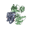
| Supramolecule | Name: Tetramer of heterodimers of SUCLG1-SUCLA2 bound to coenzyme A type: complex / ID: 1 / Parent: 0 / Macromolecule list: #1-#2 |
|---|---|
| Source (natural) | Organism:  Homo sapiens (human) Homo sapiens (human) |
| Molecular weight | Theoretical: 313 KDa |
-Macromolecule #1: Succinate--CoA ligase [ADP/GDP-forming] subunit alpha, mitochondrial
| Macromolecule | Name: Succinate--CoA ligase [ADP/GDP-forming] subunit alpha, mitochondrial type: protein_or_peptide / ID: 1 / Number of copies: 4 / Enantiomer: LEVO / EC number: succinate-CoA ligase (GDP-forming) |
|---|---|
| Source (natural) | Organism:  Homo sapiens (human) Homo sapiens (human) |
| Molecular weight | Theoretical: 32.161875 KDa |
| Recombinant expression | Organism:  |
| Sequence | String: SYTASRQHLY VDKNTKIICQ GFTGKQGTFH SQQALEYGTK LVGGTTPGKG GQTHLGLPVF NTVKEAKEQT GATASVIYVP PPFAAAAIN EAIEAEIPLV VCITEGIPQQ DMVRVKHKLL RQEKTRLIGP NCPGVINPGE CKIGIMPGHI HKKGRIGIVS R SGTLTYEA ...String: SYTASRQHLY VDKNTKIICQ GFTGKQGTFH SQQALEYGTK LVGGTTPGKG GQTHLGLPVF NTVKEAKEQT GATASVIYVP PPFAAAAIN EAIEAEIPLV VCITEGIPQQ DMVRVKHKLL RQEKTRLIGP NCPGVINPGE CKIGIMPGHI HKKGRIGIVS R SGTLTYEA VHQTTQVGLG QSLCVGIGGD PFNGTDFIDC LEIFLNDSAT EGIILIGEIG GNAEENAAEF LKQHNSGPNS KP VVSFIAG LTAPPGRRMG HAGAIIAGGK GGAKEKISAL QSAGVVVSMS PAQLGTTIYK EFEKRKML UniProtKB: Succinate--CoA ligase [ADP/GDP-forming] subunit alpha, mitochondrial |
-Macromolecule #2: Succinate--CoA ligase [ADP-forming] subunit beta, mitochondrial
| Macromolecule | Name: Succinate--CoA ligase [ADP-forming] subunit beta, mitochondrial type: protein_or_peptide / ID: 2 / Number of copies: 4 / Enantiomer: LEVO / EC number: succinate-CoA ligase (ADP-forming) |
|---|---|
| Source (natural) | Organism:  Homo sapiens (human) Homo sapiens (human) |
| Molecular weight | Theoretical: 47.757012 KDa |
| Recombinant expression | Organism:  |
| Sequence | String: MHHHHHHSSG VDLGTENLYF QSMNLSLHEY MSMELLQEAG VSVPKGYVAK SPDEAYAIAK KLGSKDVVIK AQVLAGGRGK GTFESGLKG GVKIVFSPEE AKAVSSQMIG KKLFTKQTGE KGRICNQVLV CERKYPRREY YFAITMERSF QGPVLIGSSH G GVNIEDVA ...String: MHHHHHHSSG VDLGTENLYF QSMNLSLHEY MSMELLQEAG VSVPKGYVAK SPDEAYAIAK KLGSKDVVIK AQVLAGGRGK GTFESGLKG GVKIVFSPEE AKAVSSQMIG KKLFTKQTGE KGRICNQVLV CERKYPRREY YFAITMERSF QGPVLIGSSH G GVNIEDVA AESPEAIIKE PIDIEEGIKK EQALQLAQKM GFPPNIVESA AENMVKLYSL FLKYDATMIE INPMVEDSDG AV LCMDAKI NFDSNSAYRQ KKIFDLQDWT QEDERDKDAA KANLNYIGLD GNIGCLVNGA GLAMATMDII KLHGGTPANF LDV GGGATV HQVTEAFKLI TSDKKVLAIL VNIFGGIMRC DVIAQGIVMA VKDLEIKIPV VVRLQGTRVD DAKALIADSG LKIL ACDDL DEAARMVVKL SEIVTLAKQA HVDVKFQLPI WQ UniProtKB: Succinate--CoA ligase [ADP-forming] subunit beta, mitochondrial |
-Macromolecule #3: COENZYME A
| Macromolecule | Name: COENZYME A / type: ligand / ID: 3 / Number of copies: 4 / Formula: COA |
|---|---|
| Molecular weight | Theoretical: 767.534 Da |
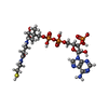
| Chemical component information |  ChemComp-COA: |
-Experimental details
-Structure determination
| Method | cryo EM |
|---|---|
 Processing Processing | single particle reconstruction |
| Aggregation state | particle |
- Sample preparation
| Concentration | 1 mg/mL | ||||||
|---|---|---|---|---|---|---|---|
| Buffer | pH: 7.5 / Component:
| ||||||
| Grid | Model: Quantifoil R1.2/1.3 / Material: GOLD / Mesh: 200 / Pretreatment - Type: GLOW DISCHARGE | ||||||
| Vitrification | Cryogen name: ETHANE / Chamber humidity: 95 % / Chamber temperature: 277.15 K / Instrument: FEI VITROBOT MARK III |
- Electron microscopy
| Microscope | TFS KRIOS |
|---|---|
| Image recording | Film or detector model: GATAN K2 QUANTUM (4k x 4k) / Detector mode: COUNTING / Number grids imaged: 1 / Number real images: 4100 / Average exposure time: 4.0 sec. / Average electron dose: 52.56 e/Å2 |
| Electron beam | Acceleration voltage: 300 kV / Electron source:  FIELD EMISSION GUN FIELD EMISSION GUN |
| Electron optics | Illumination mode: FLOOD BEAM / Imaging mode: BRIGHT FIELD / Nominal defocus max: 2.2 µm / Nominal defocus min: 1.4000000000000001 µm / Nominal magnification: 165000 |
| Sample stage | Specimen holder model: FEI TITAN KRIOS AUTOGRID HOLDER / Cooling holder cryogen: NITROGEN |
| Experimental equipment |  Model: Titan Krios / Image courtesy: FEI Company |
+ Image processing
-Atomic model buiding 1
| Initial model | PDB ID: Chain - Source name: PDB / Chain - Initial model type: experimental model |
|---|---|
| Details | Initially flexible fitting used the Namdinator server |
| Refinement | Space: REAL / Protocol: FLEXIBLE FIT |
| Output model |  PDB-9swj: |
 Movie
Movie Controller


 Z (Sec.)
 Y (Row.)
 X (Col.)


