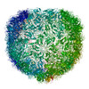
Movie
Movie Controller


+ Open data

- Basic information
| Entry | Database: PDB / ID: 9s63 | |||||||||||||||||||||||||||
|---|---|---|---|---|---|---|---|---|---|---|---|---|---|---|---|---|---|---|---|---|---|---|---|---|---|---|---|---|
| Title | Echovirus 18 in situ, virion, I4 | |||||||||||||||||||||||||||
 Components Components |
| |||||||||||||||||||||||||||
 Keywords Keywords | VIRUS / in situ structure | |||||||||||||||||||||||||||
| Function / homology |  Function and homology information Function and homology informationsymbiont-mediated suppression of host cytoplasmic pattern recognition receptor signaling pathway via inhibition of RIG-I activity / picornain 2A / symbiont-mediated suppression of host mRNA export from nucleus / symbiont genome entry into host cell via pore formation in plasma membrane / picornain 3C / T=pseudo3 icosahedral viral capsid / host cell cytoplasmic vesicle membrane / nucleoside-triphosphate phosphatase / ribonucleoside triphosphate phosphatase activity / channel activity ...symbiont-mediated suppression of host cytoplasmic pattern recognition receptor signaling pathway via inhibition of RIG-I activity / picornain 2A / symbiont-mediated suppression of host mRNA export from nucleus / symbiont genome entry into host cell via pore formation in plasma membrane / picornain 3C / T=pseudo3 icosahedral viral capsid / host cell cytoplasmic vesicle membrane / nucleoside-triphosphate phosphatase / ribonucleoside triphosphate phosphatase activity / channel activity / monoatomic ion transmembrane transport / DNA replication / RNA helicase activity / endocytosis involved in viral entry into host cell / symbiont-mediated activation of host autophagy / RNA-directed RNA polymerase / cysteine-type endopeptidase activity / viral RNA genome replication / RNA-directed RNA polymerase activity / virion attachment to host cell / host cell nucleus / structural molecule activity / DNA-templated transcription / proteolysis / RNA binding / zinc ion binding / ATP binding Similarity search - Function | |||||||||||||||||||||||||||
| Biological species |  Echovirus E18 Echovirus E18 | |||||||||||||||||||||||||||
| Method | ELECTRON MICROSCOPY / single particle reconstruction / cryo EM / Resolution: 4.3 Å | |||||||||||||||||||||||||||
 Authors Authors | Mukhamedova, L. / Plevka, P. / Trebichalska, Z. / Novacek, J. | |||||||||||||||||||||||||||
| Funding support | European Union, 1items
| |||||||||||||||||||||||||||
 Citation Citation | Journal: Proc Natl Acad Sci U S A / Year: 2026 Title: Particles of echovirus 18 open to release their genomes in vivo. Authors: Liya Mukhamedova / David Buchta / Zuzana Trebichalská / Yevgen Levdansky / Jana Moravcová / David Potěšil / Zbyněk Zdráhal / Dominik Hrebík / Lucie Nepovímová / Torleif Tollefsrud ...Authors: Liya Mukhamedova / David Buchta / Zuzana Trebichalská / Yevgen Levdansky / Jana Moravcová / David Potěšil / Zbyněk Zdráhal / Dominik Hrebík / Lucie Nepovímová / Torleif Tollefsrud Gjølberg / Jan Terje Andersen / Jiří Nováček / Tibor Füzik / Pavel Plevka /   Abstract: Enteroviruses cause a broad spectrum of human diseases, ranging from mild respiratory or gastrointestinal infections to severe neurological disorders such as aseptic meningitis and encephalitis. ...Enteroviruses cause a broad spectrum of human diseases, ranging from mild respiratory or gastrointestinal infections to severe neurological disorders such as aseptic meningitis and encephalitis. Enterovirus cell entry involves receptor-mediated endocytosis followed by destabilizing rearrangements of the virus capsid that enable genome release. However, the mechanism of enterovirus genome release has not been visualized in infected cells. Here, we used cryoelectron tomography and microscopy to image echovirus 18 (E18) entry into host cells and its interaction with the neonatal Fc receptor (FcRn). 30 min postinfection, endosomes and cytoplasm contained empty capsids missing one or several pentamers of capsid proteins, providing evidence that in vivo E18 releases its genome through capsid opening. In vitro, FcRn binding induced the expulsion of pocket factors from hydrophobic pockets in VP1, priming the virus for uncoating. The cryoelectron microscopy reconstruction of genome-containing particles of E18 inside infected cells did not reveal pocket factors, indicating that receptor binding triggers the same priming process during infection. We did not detect activated particles in infected cells, suggesting that these intermediates are short-lived and rapidly release their genomes in vivo. Our results identify capsid opening as the in vivo mechanism of echovirus 18 genome release, providing structural evidence for a process previously only inferred from in vitro experiments. | |||||||||||||||||||||||||||
| History |
|
- Structure visualization
| Structure viewer | Molecule: MolmilJmol/JSmol |
|---|
- Downloads & links
-Download
| PDBx/mmCIF format | 9s63.cif.gz | 259.3 KB | Display | PDBx/mmCIF format |
|---|---|---|---|---|
| PDB format | pdb9s63.ent.gz | 211.8 KB | Display | PDB format |
| PDBx/mmJSON format | 9s63.json.gz | Tree view | PDBx/mmJSON format | |
| Others |  Other downloads Other downloads |
-Validation report
| Arichive directory | https://data.pdbj.org/pub/pdb/validation_reports/s6/9s63ftp://data.pdbj.org/pub/pdb/validation_reports/s6/9s63 | HTTPS FTP |
|---|
-Related structure data
| Related structure data |  54622MC  9spuC  9tf0C  9tf1C  9tf2C M: map data used to model this data C: citing same article ( |
|---|---|
| Similar structure data |


-Links
 PDBj
PDBj
- Assembly
| Deposited unit | 
|
|---|---|
| 1 | x 60
|
-Components
| #1: Protein | Mass: 32564.445 Da / Num. of mol.: 1 / Source method: isolated from a natural source / Source: (natural) Echovirus E18References: UniProt: Q8V635, picornain 2A, nucleoside-triphosphate phosphatase, picornain 3C, RNA-directed RNA polymerase |
|---|---|
| #2: Protein | Mass: 28802.328 Da / Num. of mol.: 1 / Source method: isolated from a natural source / Source: (natural) Echovirus E18References: UniProt: Q8V635, picornain 2A, nucleoside-triphosphate phosphatase, picornain 3C, RNA-directed RNA polymerase |
| #3: Protein | Mass: 26143.783 Da / Num. of mol.: 1 / Source method: isolated from a natural source / Source: (natural) Echovirus E18References: UniProt: Q8V635, picornain 2A, nucleoside-triphosphate phosphatase, picornain 3C, RNA-directed RNA polymerase |
| #4: Protein | Mass: 7475.322 Da / Num. of mol.: 1 / Source method: isolated from a natural source / Source: (natural) Echovirus E18References: UniProt: Q8V635, picornain 2A, nucleoside-triphosphate phosphatase, picornain 3C, RNA-directed RNA polymerase |
| Has protein modification | N |
-Experimental details
-Experiment
| Experiment | Method: ELECTRON MICROSCOPY |
|---|---|
| EM experiment | Aggregation state: CELL / 3D reconstruction method: single particle reconstruction |
- Sample preparation
| Component | Name: Echovirus E18 / Type: VIRUS / Entity ID: all / Source: NATURAL | ||||||||||||
|---|---|---|---|---|---|---|---|---|---|---|---|---|---|
| Molecular weight |
| ||||||||||||
| Source (natural) | Organism: Echovirus E18 | ||||||||||||
| Details of virus | Empty: NO / Enveloped: NO / Isolate: OTHER / Type: VIRION | ||||||||||||
| Natural host | Organism: Homo sapiens | ||||||||||||
| Virus shell | Diameter: 330 nm | ||||||||||||
| Buffer solution | pH: 7.4 | ||||||||||||
| Specimen | Conc.: 2 mg/ml / Embedding applied: NO / Shadowing applied: NO / Staining applied: NO / Vitrification applied: YES | ||||||||||||
| Specimen support | Grid material: GOLD / Grid mesh size: 200 divisions/in. / Grid type: Quantifoil R2/1 | ||||||||||||
| Vitrification | Instrument: FEI VITROBOT MARK IV / Cryogen name: ETHANE |
- Electron microscopy imaging
| Experimental equipment |  Model: Titan Krios / Image courtesy: FEI Company |
|---|---|
| Microscopy | Model: TFS KRIOS |
| Electron gun | Electron source:  FIELD EMISSION GUN / Accelerating voltage: 300 kV / Illumination mode: FLOOD BEAM FIELD EMISSION GUN / Accelerating voltage: 300 kV / Illumination mode: FLOOD BEAM |
| Electron lens | Mode: BRIGHT FIELD / Nominal magnification: 42000 X / Nominal defocus max: 4500 nm / Nominal defocus min: 2500 nm / Alignment procedure: COMA FREE |
| Specimen holder | Cryogen: NITROGEN / Specimen holder model: FEI TITAN KRIOS AUTOGRID HOLDER |
| Image recording | Average exposure time: 8 sec. / Electron dose: 34 e/Å2 / Detector mode: COUNTING / Film or detector model: FEI FALCON III (4k x 4k) / Num. of grids imaged: 1 / Num. of real images: 1290 |
| EM imaging optics | Phase plate: OTHER |
- Processing
| EM software |
| |||||||||
|---|---|---|---|---|---|---|---|---|---|---|
| CTF correction | Type: PHASE FLIPPING AND AMPLITUDE CORRECTION | |||||||||
| Symmetry | Point symmetry: I (icosahedral) | |||||||||
| 3D reconstruction | Resolution: 4.3 Å / Resolution method: FSC 0.143 CUT-OFF / Num. of particles: 1337 / Symmetry type: POINT | |||||||||
| Atomic model building | Protocol: FLEXIBLE FIT / Space: REAL | |||||||||
| Atomic model building | PDB-ID: 6hbk Accession code: 6hbk / Source name: PDB / Type: experimental model |