Movie
Movie Controller

[English] 日本語
 Yorodumi
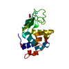
- PDB-9qum: Structure of lysozyme by continuous serial electron diffraction (... -
+ Open data

- Basic information
| Entry | Database: PDB / ID: 9qum | |||||||||
|---|---|---|---|---|---|---|---|---|---|---|
| Title | Structure of lysozyme by continuous serial electron diffraction (SerialED) | |||||||||
 Components Components | Lysozyme C | |||||||||
 Keywords Keywords | HYDROLASE / serial electron diffraction / SerialED / lysozyme | |||||||||
| Function / homology |  Function and homology information Function and homology informationLactose synthesis / Antimicrobial peptides / Neutrophil degranulation / beta-N-acetylglucosaminidase activity / cell wall macromolecule catabolic process / lysozyme / lysozyme activity / killing of cells of another organism / defense response to Gram-negative bacterium / defense response to bacterium ...Lactose synthesis / Antimicrobial peptides / Neutrophil degranulation / beta-N-acetylglucosaminidase activity / cell wall macromolecule catabolic process / lysozyme / lysozyme activity / killing of cells of another organism / defense response to Gram-negative bacterium / defense response to bacterium / defense response to Gram-positive bacterium / endoplasmic reticulum / : / identical protein binding / cytoplasm Similarity search - Function | |||||||||
| Biological species |  | |||||||||
| Method | ELECTRON CRYSTALLOGRAPHY / electron crystallography / cryo EM / Resolution: 0.83 Å | |||||||||
 Authors Authors | Hofer, G. / Wang, L. / Pacoste, L. / Hager, P. / Fonjallaz, A. / Scaletti Hutchinson, E. / Stenmark, P. / Di Palma, M. / Williams, L. / Worral, J. ...Hofer, G. / Wang, L. / Pacoste, L. / Hager, P. / Fonjallaz, A. / Scaletti Hutchinson, E. / Stenmark, P. / Di Palma, M. / Williams, L. / Worral, J. / Steiner, R. / Xu, H. / Zou, X. | |||||||||
| Funding support |  Sweden, 2items Sweden, 2items
| |||||||||
 Citation Citation | Journal: To Be Published Title: Continuous Serial Electron Diffraction for High Quality Protein Structures Authors: Hofer, G. / Wang, L. / Pacoste, L. / Hager, P. / Fonjallaz, A. / Williams, L. / Worrall, J. / Steiner, R. / Xu, H. / Zou, X. | |||||||||
| History |
|
- Structure visualization
| Structure viewer | Molecule: MolmilJmol/JSmol |
|---|
- Downloads & links
-Download
| PDBx/mmCIF format | 9qum.cif.gz | 114.3 KB | Display | PDBx/mmCIF format |
|---|---|---|---|---|
| PDB format | pdb9qum.ent.gz | 72.1 KB | Display | PDB format |
| PDBx/mmJSON format | 9qum.json.gz | Tree view | PDBx/mmJSON format | |
| Others |  Other downloads Other downloads |
-Validation report
| Arichive directory | https://data.pdbj.org/pub/pdb/validation_reports/qu/9qumftp://data.pdbj.org/pub/pdb/validation_reports/qu/9qum | HTTPS FTP |
|---|
-Related structure data
| Related structure data |  9queC  9quhC  9qukC  9qw5C  9qw6C C: citing same article ( |
|---|---|
| Similar structure data | |
| Experimental dataset #1 | Data reference: 10.15785/SBGRID/1150 / Data set type: diffraction image data |
-Links
 PDBj
PDBj


- Assembly
| Deposited unit | 
| ||||||||||||
|---|---|---|---|---|---|---|---|---|---|---|---|---|---|
| 1 |
| ||||||||||||
| Unit cell |
|
-Components
| #1: Protein | Mass: 16257.660 Da / Num. of mol.: 1 / Source method: isolated from a natural source / Source: (natural) | ||||||
|---|---|---|---|---|---|---|---|
| #2: Chemical | ChemComp-ACT /   Mass: 59.044 Da / Num. of mol.: 1 / Source method: obtained synthetically / Formula: C2H3O2 Mass: 59.044 Da / Num. of mol.: 1 / Source method: obtained synthetically / Formula: C2H3O2 | ||||||
| #3: Chemical |   Mass: 35.453 Da / Num. of mol.: 3 / Source method: obtained synthetically / Formula: Cl Mass: 35.453 Da / Num. of mol.: 3 / Source method: obtained synthetically / Formula: Cl#4: Water | ChemComp-HOH / |  Mass: 18.015 Da / Num. of mol.: 147 / Source method: isolated from a natural source / Formula: H2O Mass: 18.015 Da / Num. of mol.: 147 / Source method: isolated from a natural source / Formula: H2OHas ligand of interest | N | Has protein modification | Y | |
-Experimental details
-Experiment
| Experiment | Method: ELECTRON CRYSTALLOGRAPHY |
|---|---|
| EM experiment | Aggregation state: 3D ARRAY / 3D reconstruction method: electron crystallography |
- Sample preparation
| Component | Name: Lysozyme / Type: ORGANELLE OR CELLULAR COMPONENT / Entity ID: #1 / Source: NATURAL | |||||||||||||||
|---|---|---|---|---|---|---|---|---|---|---|---|---|---|---|---|---|
| Molecular weight | Value: 0.0162 MDa / Experimental value: NO | |||||||||||||||
| Source (natural) | Organism: | |||||||||||||||
| Buffer solution | pH: 4.5 Details: Crystals were produced by adding 1 part of lysozyme solution (40 mg/mL) to 1 part of precipitant (0.8 M NaNO3, 50mM NaAc, pH 4.5) | |||||||||||||||
| Buffer component |
| |||||||||||||||
| Specimen | Conc.: 40 mg/ml / Embedding applied: NO / Shadowing applied: NO / Staining applied: NO / Vitrification applied: YES / Details: Hen egg white lysozyme | |||||||||||||||
| Specimen support | Grid material: COPPER / Grid mesh size: 200 divisions/in. / Grid type: Quantifoil R0.6/1 | |||||||||||||||
| Vitrification | Cryogen name: ETHANE Details: Manual blotting in room temperature with ambient humidity |
-Data collection
| Experimental equipment |  Model: Titan Krios / Image courtesy: FEI Company | |||||||||||||||||||||
|---|---|---|---|---|---|---|---|---|---|---|---|---|---|---|---|---|---|---|---|---|---|---|
| Microscopy | Model: TFS KRIOS | |||||||||||||||||||||
| Electron gun | Electron source:  FIELD EMISSION GUN / Accelerating voltage: 300 kV / Illumination mode: FLOOD BEAM FIELD EMISSION GUN / Accelerating voltage: 300 kV / Illumination mode: FLOOD BEAM | |||||||||||||||||||||
| Electron lens | Mode: DIFFRACTION / Nominal defocus max: 0 nm / Nominal defocus min: 0 nm / Calibrated defocus min: 0 nm / C2 aperture diameter: 20 µm | |||||||||||||||||||||
| Specimen holder | Cryogen: NITROGEN / Specimen holder model: FEI TITAN KRIOS AUTOGRID HOLDER / Temperature (max): 98 K / Temperature (min): 78 K | |||||||||||||||||||||
| Image recording | Electron dose: 1.52 e/Å2 / Film or detector model: FEI CETA (4k x 4k) / Details: FEI Ceta-D CMOS detector | |||||||||||||||||||||
| EM diffraction shell |
| |||||||||||||||||||||
| EM diffraction stats | Fourier space coverage: 99.9 % / High resolution: 0.83 Å / Num. of intensities measured: 5003763 / Num. of structure factors: 89374 / Phase error rejection criteria: 0 / Rmerge: 0.109 | |||||||||||||||||||||
| Reflection | Biso Wilson estimate: 8.7 Å2 |
- Processing
| EM software |
| |||||||||||||||||||||||||||||||||||||||||||||||||||||||||||||||||||||||||||||||||||||||||||||||||||||||||
|---|---|---|---|---|---|---|---|---|---|---|---|---|---|---|---|---|---|---|---|---|---|---|---|---|---|---|---|---|---|---|---|---|---|---|---|---|---|---|---|---|---|---|---|---|---|---|---|---|---|---|---|---|---|---|---|---|---|---|---|---|---|---|---|---|---|---|---|---|---|---|---|---|---|---|---|---|---|---|---|---|---|---|---|---|---|---|---|---|---|---|---|---|---|---|---|---|---|---|---|---|---|---|---|---|---|---|
| EM 3D crystal entity | ∠α: 87.73 ° / ∠β: 108.97 ° / ∠γ: 111.6 ° / A: 26.66 Å / B: 31.15 Å / C: 33.57 Å / Space group name: P1 / Space group num: 1 | |||||||||||||||||||||||||||||||||||||||||||||||||||||||||||||||||||||||||||||||||||||||||||||||||||||||||
| CTF correction | Type: NONE | |||||||||||||||||||||||||||||||||||||||||||||||||||||||||||||||||||||||||||||||||||||||||||||||||||||||||
| 3D reconstruction | Resolution: 0.83 Å / Resolution method: DIFFRACTION PATTERN/LAYERLINES / Symmetry type: 3D CRYSTAL | |||||||||||||||||||||||||||||||||||||||||||||||||||||||||||||||||||||||||||||||||||||||||||||||||||||||||
| Atomic model building | B value: 11.04 / Protocol: OTHER / Space: RECIPROCAL / Target criteria: maximum-likelihood | |||||||||||||||||||||||||||||||||||||||||||||||||||||||||||||||||||||||||||||||||||||||||||||||||||||||||
| Atomic model building | PDB-ID: 7SKW Accession code: 7SKW / Source name: PDB / Type: experimental model | |||||||||||||||||||||||||||||||||||||||||||||||||||||||||||||||||||||||||||||||||||||||||||||||||||||||||
| Refinement | Resolution: 0.83→11.73 Å / SU ML: 0.1562 / Cross valid method: FREE R-VALUE / σ(F): 1.96 / Phase error: 28.8099 Stereochemistry target values: GeoStd + Monomer Library + CDL v1.2
| |||||||||||||||||||||||||||||||||||||||||||||||||||||||||||||||||||||||||||||||||||||||||||||||||||||||||
| Solvent computation | Shrinkage radii: 0.9 Å / VDW probe radii: 1.11 Å / Solvent model: FLAT BULK SOLVENT MODEL | |||||||||||||||||||||||||||||||||||||||||||||||||||||||||||||||||||||||||||||||||||||||||||||||||||||||||
| Displacement parameters | Biso mean: 11.04 Å2 | |||||||||||||||||||||||||||||||||||||||||||||||||||||||||||||||||||||||||||||||||||||||||||||||||||||||||
| Refinement step | Cycle: LAST / Resolution: 0.83→11.73 Å
| |||||||||||||||||||||||||||||||||||||||||||||||||||||||||||||||||||||||||||||||||||||||||||||||||||||||||
| Refine LS restraints |
| |||||||||||||||||||||||||||||||||||||||||||||||||||||||||||||||||||||||||||||||||||||||||||||||||||||||||
| LS refinement shell |
|