Movie
Movie Controller

[English] 日本語
 Yorodumi
- PDB-9quk: Structure of human MTH1 in complex with 8DG by MicroED using low ... -
+ Open data

- Basic information
| Entry | Database: PDB / ID: 9quk | |||||||||
|---|---|---|---|---|---|---|---|---|---|---|
| Title | Structure of human MTH1 in complex with 8DG by MicroED using low electron fluence | |||||||||
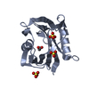
 Components Components | 7,8-dihydro-8-oxoguanine triphosphatase | |||||||||
 Keywords Keywords | HYDROLASE / serial electron diffraction / SerialED / MicroED / MTH1 / 8DG | |||||||||
| Function / homology |  Function and homology information Function and homology information2-hydroxy-ATP hydrolase activity / 2-hydroxy-dATP hydrolase activity / N6-methyl-(d)ATP hydrolase activity / O6-methyl-dGTP hydrolase activity / 2-hydroxy-dATP diphosphatase / dATP diphosphatase activity / ATP diphosphatase activity / 8-oxo-7,8-dihydrodeoxyguanosine triphosphate pyrophosphatase activity / 8-oxo-7,8-dihydroguanosine triphosphate pyrophosphatase activity / DNA protection ...2-hydroxy-ATP hydrolase activity / 2-hydroxy-dATP hydrolase activity / N6-methyl-(d)ATP hydrolase activity / O6-methyl-dGTP hydrolase activity / 2-hydroxy-dATP diphosphatase / dATP diphosphatase activity / ATP diphosphatase activity / 8-oxo-7,8-dihydrodeoxyguanosine triphosphate pyrophosphatase activity / 8-oxo-7,8-dihydroguanosine triphosphate pyrophosphatase activity / DNA protection / Phosphate bond hydrolysis by NUDT proteins / hydrolase activity, acting on acid anhydrides, in phosphorus-containing anhydrides / purine nucleoside catabolic process / snoRNA binding / Hydrolases; Acting on acid anhydrides; In phosphorus-containing anhydrides / response to oxidative stress / mitochondrial matrix / DNA repair / mitochondrion / metal ion binding / nucleus / cytoplasm / cytosol Similarity search - Function | |||||||||
| Biological species |  Homo sapiens (human) Homo sapiens (human) | |||||||||
| Method | ELECTRON CRYSTALLOGRAPHY / electron crystallography / cryo EM / Resolution: 2.86 Å | |||||||||
 Authors Authors | Hofer, G. / Wang, L. / Pacoste, L. / Hager, P. / Fonjallaz, A. / Scaletti Hutchinson, E. / Stenmark, P. / Di Palma, M. / Williams, L. / Worral, J. ...Hofer, G. / Wang, L. / Pacoste, L. / Hager, P. / Fonjallaz, A. / Scaletti Hutchinson, E. / Stenmark, P. / Di Palma, M. / Williams, L. / Worral, J. / Steiner, R. / Xu, H. / Zou, X. | |||||||||
| Funding support |  Sweden, 2items Sweden, 2items
| |||||||||
 Citation Citation | Journal: To Be Published Title: Continuous Serial Electron Diffraction for High Quality Protein Structures Authors: Hofer, G. / Wang, L. / Pacoste, L. / Hager, P. / Fonjallaz, A. / Williams, L. / Worrall, J. / Steiner, R. / Xu, H. / Zou, X. | |||||||||
| History |
|
- Structure visualization
| Structure viewer | Molecule: MolmilJmol/JSmol |
|---|
- Downloads & links
-Download
| PDBx/mmCIF format | 9quk.cif.gz | 152.1 KB | Display | PDBx/mmCIF format |
|---|---|---|---|---|
| PDB format | pdb9quk.ent.gz | 100.4 KB | Display | PDB format |
| PDBx/mmJSON format | 9quk.json.gz | Tree view | PDBx/mmJSON format | |
| Others |  Other downloads Other downloads |
-Validation report
| Arichive directory | https://data.pdbj.org/pub/pdb/validation_reports/qu/9qukftp://data.pdbj.org/pub/pdb/validation_reports/qu/9quk | HTTPS FTP |
|---|
-Related structure data
| Related structure data |  9queC  9quhC  9qumC  9qw5C  9qw6C C: citing same article ( |
|---|---|
| Similar structure data | |
| Experimental dataset #1 | Data reference: 10.5281/zenodo.15168593 / Data set type: diffraction image data |
-Links
 PDBj
PDBj

- Assembly
| Deposited unit | 
| ||||||||||||
|---|---|---|---|---|---|---|---|---|---|---|---|---|---|
| 1 | 
| ||||||||||||
| 2 | 
| ||||||||||||
| Unit cell |
|
-Components
| #1: Protein | Mass: 18253.736 Da / Num. of mol.: 2 Source method: isolated from a genetically manipulated source Source: (gene. exp.) Homo sapiens (human) / Gene: NUDT1, MTH1 / Production host:  References: UniProt: P36639, 8-oxo-dGTP diphosphatase, 2-hydroxy-dATP diphosphatase #2: Chemical | 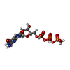  Mass: 523.180 Da / Num. of mol.: 2 / Source method: obtained synthetically / Formula: C10H16N5O14P3 / Feature type: SUBJECT OF INVESTIGATION Mass: 523.180 Da / Num. of mol.: 2 / Source method: obtained synthetically / Formula: C10H16N5O14P3 / Feature type: SUBJECT OF INVESTIGATION#3: Chemical | ChemComp-SO4 /   Mass: 96.063 Da / Num. of mol.: 5 / Source method: obtained synthetically / Formula: SO4 Mass: 96.063 Da / Num. of mol.: 5 / Source method: obtained synthetically / Formula: SO4#4: Water | ChemComp-HOH / |  Mass: 18.015 Da / Num. of mol.: 11 / Source method: isolated from a natural source / Formula: H2O Mass: 18.015 Da / Num. of mol.: 11 / Source method: isolated from a natural source / Formula: H2OHas ligand of interest | Y | Has protein modification | N | |
|---|
-Experimental details
-Experiment
| Experiment | Method: ELECTRON CRYSTALLOGRAPHY |
|---|---|
| EM experiment | Aggregation state: 3D ARRAY / 3D reconstruction method: electron crystallography |
- Sample preparation
| Component | Name: 7,8-dihydro-8-oxoguanine triphosphatase / Type: ORGANELLE OR CELLULAR COMPONENT / Entity ID: #1 / Source: RECOMBINANT |
|---|---|
| Molecular weight | Value: 0.0182 MDa / Experimental value: NO |
| Source (natural) | Organism: Homo sapiens (human) |
| Source (recombinant) | Organism: |
| Buffer solution | pH: 4 |
| Specimen | Conc.: 14 mg/ml / Embedding applied: NO / Shadowing applied: NO / Staining applied: NO / Vitrification applied: YES |
| Specimen support | Grid material: COPPER / Grid mesh size: 200 divisions/in. / Grid type: C-flat-1.2/1.3 |
| Vitrification | Cryogen name: ETHANE Details: Manual blotting at room temperature with ambient humidity |
-Data collection
| Experimental equipment |  Model: Titan Krios / Image courtesy: FEI Company | |||||||||||||||||||||
|---|---|---|---|---|---|---|---|---|---|---|---|---|---|---|---|---|---|---|---|---|---|---|
| Microscopy | Model: TFS KRIOS | |||||||||||||||||||||
| Electron gun | Electron source:  FIELD EMISSION GUN / Accelerating voltage: 300 kV / Illumination mode: FLOOD BEAM FIELD EMISSION GUN / Accelerating voltage: 300 kV / Illumination mode: FLOOD BEAM | |||||||||||||||||||||
| Electron lens | Mode: DIFFRACTION / Nominal defocus max: 0 nm / Nominal defocus min: 0 nm / Calibrated defocus min: 0 nm / Alignment procedure: BASIC | |||||||||||||||||||||
| Specimen holder | Cryogen: NITROGEN / Specimen holder model: FEI TITAN KRIOS AUTOGRID HOLDER / Temperature (max): 98 K / Temperature (min): 78 K | |||||||||||||||||||||
| Image recording | Electron dose: 0.055 e/Å2 / Film or detector model: FEI CETA (4k x 4k) / Details: FEI Ceta-D CMOS detector | |||||||||||||||||||||
| EM diffraction shell |
| |||||||||||||||||||||
| EM diffraction stats | Fourier space coverage: 85.3 % / High resolution: 2.86 Å / Num. of intensities measured: 127640 / Num. of structure factors: 6638 / Phase error rejection criteria: 0 / Rmerge: 54.3 | |||||||||||||||||||||
| Reflection | Biso Wilson estimate: 33.69 Å2 |
- Processing
| Image processing | Details: FEI Ceta-D CMOS detector | |||||||||||||||||||||||||||||||||||
|---|---|---|---|---|---|---|---|---|---|---|---|---|---|---|---|---|---|---|---|---|---|---|---|---|---|---|---|---|---|---|---|---|---|---|---|---|
| EM 3D crystal entity | ∠α: 90 ° / ∠β: 90 ° / ∠γ: 90 ° / A: 59.34 Å / B: 67.55 Å / C: 80.11 Å / Space group name: P212121 / Space group num: 19 | |||||||||||||||||||||||||||||||||||
| CTF correction | Type: NONE | |||||||||||||||||||||||||||||||||||
| 3D reconstruction | Resolution: 2.86 Å / Resolution method: DIFFRACTION PATTERN/LAYERLINES / Symmetry type: 3D CRYSTAL | |||||||||||||||||||||||||||||||||||
| Atomic model building | B value: 23.9 / Protocol: OTHER / Target criteria: maxiumum likelihood | |||||||||||||||||||||||||||||||||||
| Atomic model building | PDB-ID: 3ZR1 Accession code: 3ZR1 / Source name: PDB / Type: experimental model | |||||||||||||||||||||||||||||||||||
| Refinement | Resolution: 2.86→29.67 Å / SU ML: 0.2754 / Cross valid method: FREE R-VALUE / σ(F): 1.34 / Phase error: 27.9872 Stereochemistry target values: GeoStd + Monomer Library + CDL v1.2
| |||||||||||||||||||||||||||||||||||
| Solvent computation | Shrinkage radii: 0.9 Å / VDW probe radii: 1.1 Å / Solvent model: FLAT BULK SOLVENT MODEL | |||||||||||||||||||||||||||||||||||
| Displacement parameters | Biso mean: 22.6 Å2 | |||||||||||||||||||||||||||||||||||
| Refine LS restraints |
| |||||||||||||||||||||||||||||||||||
| LS refinement shell |
|