 Movie
Movie Controller


+ Open data

- Basic information
| Entry | Database: PDB / ID: 9qgk | |||||||||||||||||||||||||||
|---|---|---|---|---|---|---|---|---|---|---|---|---|---|---|---|---|---|---|---|---|---|---|---|---|---|---|---|---|
| Title | F-actin decorated by ITPKA | |||||||||||||||||||||||||||
 Components Components |
| |||||||||||||||||||||||||||
 Keywords Keywords | CYTOSOLIC PROTEIN / actin | |||||||||||||||||||||||||||
| Function / homology |  Function and homology information Function and homology informationinositol-trisphosphate 3-kinase / inositol-1,4,5-trisphosphate 3-kinase activity / inositol hexakisphosphate kinase activity / inositol phosphate biosynthetic process / modification of postsynaptic actin cytoskeleton / postsynaptic actin cytoskeleton / calcium/calmodulin-dependent protein kinase activity / phosphatidylinositol phosphate biosynthetic process / Synthesis of IP3 and IP4 in the cytosol / striated muscle thin filament ...inositol-trisphosphate 3-kinase / inositol-1,4,5-trisphosphate 3-kinase activity / inositol hexakisphosphate kinase activity / inositol phosphate biosynthetic process / modification of postsynaptic actin cytoskeleton / postsynaptic actin cytoskeleton / calcium/calmodulin-dependent protein kinase activity / phosphatidylinositol phosphate biosynthetic process / Synthesis of IP3 and IP4 in the cytosol / striated muscle thin filament / skeletal muscle thin filament assembly / skeletal muscle fiber development / stress fiber / cellular response to calcium ion / actin filament / response to calcium ion / Hydrolases; Acting on acid anhydrides; Acting on acid anhydrides to facilitate cellular and subcellular movement / small GTPase binding / structural constituent of cytoskeleton / actin cytoskeleton / dendritic spine / cytoskeleton / calmodulin binding / hydrolase activity / glutamatergic synapse / signal transduction / ATP binding / nucleus / cytosol / cytoplasm Similarity search - Function | |||||||||||||||||||||||||||
| Biological species |  Homo sapiens (human) Homo sapiens (human)  Gallus (bird) Gallus (bird) Amanita phalloides (death cap) Amanita phalloides (death cap) | |||||||||||||||||||||||||||
| Method | ELECTRON MICROSCOPY / helical reconstruction / cryo EM / Resolution: 2.97 Å | |||||||||||||||||||||||||||
 Authors Authors | Yuan, B. / Paraschiakos, T. / Windhorst, S. / Marlovits, T.C. | |||||||||||||||||||||||||||
| Funding support | 1items
| |||||||||||||||||||||||||||
 Citation Citation | Journal: Nat Cell Biol / Year: 2026 Title: Evolutionarily conserved short linear motifs drive actin filament binding. Authors: Themistoklis Paraschiakos / Biao Yuan / Michael Hecht-Bucher / Kostiantyn Sopelniak / Pasquale Cervero / Lisa Simon / Ksenija Zonjic / Dominic Eggers / Franziska Selle / Jing Li / Ali ...Authors: Themistoklis Paraschiakos / Biao Yuan / Michael Hecht-Bucher / Kostiantyn Sopelniak / Pasquale Cervero / Lisa Simon / Ksenija Zonjic / Dominic Eggers / Franziska Selle / Jing Li / Ali Biabani / Stefan Linder / Thomas C Marlovits / Sabine Windhorst /  Abstract: Regulation of the actin cytoskeleton by actin-binding proteins is essential for cellular homeostasis, and the mode of actin binding determines the activity of actin-binding proteins. Here we identify ...Regulation of the actin cytoskeleton by actin-binding proteins is essential for cellular homeostasis, and the mode of actin binding determines the activity of actin-binding proteins. Here we identify a 'short linear actin filament-binding motif' (SFM) based on the cryo-electron microscopy structure of the ITPKA-actin filament complex. Using the computational pipeline SLiMFold, we discovered 103 human proteins containing SFMs with diverse cellular roles. Phylogenetic analysis suggests that SFMs arose de novo and are conserved across eukaryotes, exhibiting actin filament-binding affinities of 2-12 µM. Critical residues mediating binding and modulating affinity were defined, and the cryo-electron microscopy structures of two SFM-actin filament complexes revealed that SFM binding decreases actin-filament stiffness. These findings indicate that SFMs regulate actin-filament conformation and serve as anchoring modules that connect actin dynamics to a broad variety of cellular functions, providing a framework for understanding the actin-associated roles of numerous proteins. | |||||||||||||||||||||||||||
| History |
|
- Structure visualization
| Structure viewer | Molecule: MolmilJmol/JSmol |
|---|
- Downloads & links
-Download
| PDBx/mmCIF format | 9qgk.cif.gz | 793.7 KB | Display | PDBx/mmCIF format |
|---|---|---|---|---|
| PDB format | pdb9qgk.ent.gz | 543.4 KB | Display | PDB format |
| PDBx/mmJSON format | 9qgk.json.gz | Tree view | PDBx/mmJSON format | |
| Others |  Other downloads Other downloads |
-Validation report
| Arichive directory | https://data.pdbj.org/pub/pdb/validation_reports/qg/9qgkftp://data.pdbj.org/pub/pdb/validation_reports/qg/9qgk | HTTPS FTP |
|---|
-Related structure data
| Related structure data |  53133MC 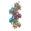 8r3hC  9sgkC M: map data used to model this data C: citing same article ( |
|---|---|
| Similar structure data |

-Links
 PDBj
PDBj





- Assembly
| Deposited unit | 
|
|---|---|
| 1 |
|
-Components
| #1: Protein | Mass: 51083.820 Da / Num. of mol.: 5 Source method: isolated from a genetically manipulated source Source: (gene. exp.) Homo sapiens (human) / Gene: ITPKAProduction host:   Escherichia coli 'BL21-Gold(DE3)pLysS AG' (bacteria) Escherichia coli 'BL21-Gold(DE3)pLysS AG' (bacteria)References: UniProt: P23677 #2: Protein | Mass: 42109.973 Da / Num. of mol.: 5 Source method: isolated from a genetically manipulated source Source: (gene. exp.) Gallus (bird) / Gene: ACTA1, ACTA / Production host: Gallus (bird)References: UniProt: P68139, Hydrolases; Acting on acid anhydrides; Acting on acid anhydrides to facilitate cellular and subcellular movement #3: Protein/peptide | Mass: 808.899 Da / Num. of mol.: 3 Source method: isolated from a genetically manipulated source Source: (gene. exp.) Amanita phalloides (death cap) / Production host: Amanita phalloides (death cap)#4: Chemical | ChemComp-MG /   Mass: 24.305 Da / Num. of mol.: 5 / Source method: obtained synthetically / Formula: Mg / Feature type: SUBJECT OF INVESTIGATION Mass: 24.305 Da / Num. of mol.: 5 / Source method: obtained synthetically / Formula: Mg / Feature type: SUBJECT OF INVESTIGATION#5: Chemical | ChemComp-ADP /  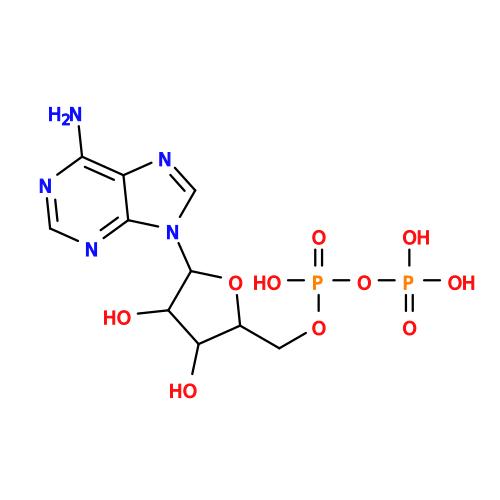 Mass: 427.201 Da / Num. of mol.: 5 / Source method: obtained synthetically / Formula: C10H15N5O10P2 / Feature type: SUBJECT OF INVESTIGATION / Comment: ADP, energy-carrying molecule*YM Mass: 427.201 Da / Num. of mol.: 5 / Source method: obtained synthetically / Formula: C10H15N5O10P2 / Feature type: SUBJECT OF INVESTIGATION / Comment: ADP, energy-carrying molecule*YMHas ligand of interest | Y | Has protein modification | Y | |
|---|
-Experimental details
-Experiment
| Experiment | Method: ELECTRON MICROSCOPY |
|---|---|
| EM experiment | Aggregation state: FILAMENT / 3D reconstruction method: helical reconstruction |
- Sample preparation
| Component | Name: F-actin decorated by ITPKA / Type: COMPLEX / Entity ID: #1-#3 / Source: RECOMBINANT |
|---|---|
| Source (natural) | Organism: Homo sapiens (human) |
| Source (recombinant) | Organism: Escherichia coli 'BL21-Gold(DE3)pLysS AG' (bacteria) |
| Buffer solution | pH: 8 |
| Specimen | Embedding applied: NO / Shadowing applied: NO / Staining applied: NO / Vitrification applied: YES |
| Vitrification | Cryogen name: ETHANE-PROPANE |
- Electron microscopy imaging
| Experimental equipment |  Model: Titan Krios / Image courtesy: FEI Company |
|---|---|
| Microscopy | Model: TFS KRIOS |
| Electron gun | Electron source:  FIELD EMISSION GUN / Accelerating voltage: 300 kV / Illumination mode: FLOOD BEAM FIELD EMISSION GUN / Accelerating voltage: 300 kV / Illumination mode: FLOOD BEAM |
| Electron lens | Mode: BRIGHT FIELD / Nominal defocus max: 3000 nm / Nominal defocus min: 800 nm |
| Image recording | Electron dose: 60 e/Å2 / Film or detector model: GATAN K3 (6k x 4k) |
- Processing
| CTF correction | Type: PHASE FLIPPING AND AMPLITUDE CORRECTION | ||||||||||||||||||||||||
|---|---|---|---|---|---|---|---|---|---|---|---|---|---|---|---|---|---|---|---|---|---|---|---|---|---|
| Helical symmerty | Angular rotation/subunit: -166.86 ° / Axial rise/subunit: 27.58 Å / Axial symmetry: C1 | ||||||||||||||||||||||||
| 3D reconstruction | Resolution: 2.97 Å / Resolution method: FSC 0.143 CUT-OFF / Num. of particles: 65917 / Symmetry type: HELICAL | ||||||||||||||||||||||||
| Refinement | Cross valid method: NONE Stereochemistry target values: GeoStd + Monomer Library + CDL v1.2 | ||||||||||||||||||||||||
| Displacement parameters | Biso mean: 44.11 Å2 | ||||||||||||||||||||||||
| Refine LS restraints |
|
