 Movie
Movie Controller


+ Open data

- Basic information
| Entry |  | |||||||||
|---|---|---|---|---|---|---|---|---|---|---|
| Title | F-actin in complex with USP54 M1 actin binding motif | |||||||||
 Map data Map data | ||||||||||
 Sample Sample |
| |||||||||
 Keywords Keywords | Actin / STRUCTURAL PROTEIN | |||||||||
| Function / homology |  Function and homology information Function and homology informationstriated muscle thin filament / skeletal muscle thin filament assembly / skeletal muscle fiber development / stress fiber / actin filament / Hydrolases; Acting on acid anhydrides; Acting on acid anhydrides to facilitate cellular and subcellular movement / structural constituent of cytoskeleton / actin cytoskeleton / hydrolase activity / ATP binding Similarity search - Function | |||||||||
| Biological species |  Homo sapiens (human) / Homo sapiens (human) /  | |||||||||
| Method | helical reconstruction / cryo EM / Resolution: 3.8 Å | |||||||||
 Authors Authors | Yuan B / Paraschiakos T / Windhorst S / Marlovits TC | |||||||||
| Funding support | 1 items
| |||||||||
 Citation Citation | Journal: Nat Cell Biol / Year: 2026 Title: Evolutionarily conserved short linear motifs drive actin filament binding. Authors: Themistoklis Paraschiakos / Biao Yuan / Michael Hecht-Bucher / Kostiantyn Sopelniak / Pasquale Cervero / Lisa Simon / Ksenija Zonjic / Dominic Eggers / Franziska Selle / Jing Li / Ali ...Authors: Themistoklis Paraschiakos / Biao Yuan / Michael Hecht-Bucher / Kostiantyn Sopelniak / Pasquale Cervero / Lisa Simon / Ksenija Zonjic / Dominic Eggers / Franziska Selle / Jing Li / Ali Biabani / Stefan Linder / Thomas C Marlovits / Sabine Windhorst /  Abstract: Regulation of the actin cytoskeleton by actin-binding proteins is essential for cellular homeostasis, and the mode of actin binding determines the activity of actin-binding proteins. Here we identify ...Regulation of the actin cytoskeleton by actin-binding proteins is essential for cellular homeostasis, and the mode of actin binding determines the activity of actin-binding proteins. Here we identify a 'short linear actin filament-binding motif' (SFM) based on the cryo-electron microscopy structure of the ITPKA-actin filament complex. Using the computational pipeline SLiMFold, we discovered 103 human proteins containing SFMs with diverse cellular roles. Phylogenetic analysis suggests that SFMs arose de novo and are conserved across eukaryotes, exhibiting actin filament-binding affinities of 2-12 µM. Critical residues mediating binding and modulating affinity were defined, and the cryo-electron microscopy structures of two SFM-actin filament complexes revealed that SFM binding decreases actin-filament stiffness. These findings indicate that SFMs regulate actin-filament conformation and serve as anchoring modules that connect actin dynamics to a broad variety of cellular functions, providing a framework for understanding the actin-associated roles of numerous proteins. | |||||||||
| History |
|
- Structure visualization
| Supplemental images |
|---|

- Downloads & links
-EMDB archive
| Map data | emd_54871.map.gz | 59.5 MB | EMDB map data format | |
|---|---|---|---|---|
| Header (meta data) | emd-54871-v30.xmlemd-54871.xml | 20.2 KB 20.2 KB | Display Display | EMDB header |
| FSC (resolution estimation) | emd_54871_fsc.xml | 8.5 KB | Display | FSC data file |
| Images |  emd_54871.png emd_54871.png | 69.9 KB | ||
| Filedesc metadata | emd-54871.cif.gz | 6.6 KB | ||
| Others | emd_54871_half_map_1.map.gzemd_54871_half_map_2.map.gz | 59.4 MB 59.4 MB | ||
| Archive directory |  http://ftp.pdbj.org/pub/emdb/structures/EMD-54871ftp://ftp.pdbj.org/pub/emdb/structures/EMD-54871 http://ftp.pdbj.org/pub/emdb/structures/EMD-54871ftp://ftp.pdbj.org/pub/emdb/structures/EMD-54871 | HTTPS FTP |
-Related structure data
| Related structure data |  9sgkMC 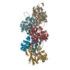 8r3hC  9qgkC M: atomic model generated by this map C: citing same article ( |
|---|---|
| Similar structure data |

-Links
| EMDB pages | EMDB (EBI/PDBe) / EMDataResource |
|---|---|
| Related items in Molecule of the Month |


-Map
| File | Download / File: emd_54871.map.gz / Format: CCP4 / Size: 64 MB / Type: IMAGE STORED AS FLOATING POINT NUMBER (4 BYTES) | ||||||||||||||||||||||||||||||||||||
|---|---|---|---|---|---|---|---|---|---|---|---|---|---|---|---|---|---|---|---|---|---|---|---|---|---|---|---|---|---|---|---|---|---|---|---|---|---|
| Projections & slices | Image control
Images are generated by Spider. | ||||||||||||||||||||||||||||||||||||
| Voxel size | X=Y=Z: 0.91 Å | ||||||||||||||||||||||||||||||||||||
| Density |
| ||||||||||||||||||||||||||||||||||||
| Symmetry | Space group: 1 | ||||||||||||||||||||||||||||||||||||
| Details | EMDB XML:
|
 Z (Sec.)
 Y (Row.)
 X (Col.)


-Supplemental data
-Half map: #2
| File | emd_54871_half_map_1.map | ||||||||||||
|---|---|---|---|---|---|---|---|---|---|---|---|---|---|
| Projections & Slices |
| ||||||||||||
| Density Histograms |


-Half map: #1
| File | emd_54871_half_map_2.map | ||||||||||||
|---|---|---|---|---|---|---|---|---|---|---|---|---|---|
| Projections & Slices |
| ||||||||||||
| Density Histograms |


- Sample components
-Entire : F-actin-USP54_M1
| Entire | Name: F-actin-USP54_M1 |
|---|---|
| Components |
|
-Supramolecule #1: F-actin-USP54_M1
| Supramolecule | Name: F-actin-USP54_M1 / type: complex / ID: 1 / Parent: 0 / Macromolecule list: #1-#2 |
|---|---|
| Source (natural) | Organism: Homo sapiens (human) |
-Macromolecule #1: Actin binding motif
| Macromolecule | Name: Actin binding motif / type: protein_or_peptide / ID: 1 / Number of copies: 5 / Enantiomer: LEVO |
|---|---|
| Source (natural) | Organism: Homo sapiens (human) |
| Molecular weight | Theoretical: 33.529469 KDa |
| Recombinant expression | Organism:  |
| Sequence | String: MIPSTKGSVR SLIEQFERIQ GVSAGDPPVA TSVSKGEELF TGVVPILVEL DGDVNGHKFS VSGEGEGDAT YGKLTLKFIC TTGKLPVPW PTLVTTLTYG VQCFSRYPDH MKQHDFFKSA MPEGYVQERT IFFKDDGNYK TRAEVKFEGD TLVNRIELKG I DFKEDGNI ...String: MIPSTKGSVR SLIEQFERIQ GVSAGDPPVA TSVSKGEELF TGVVPILVEL DGDVNGHKFS VSGEGEGDAT YGKLTLKFIC TTGKLPVPW PTLVTTLTYG VQCFSRYPDH MKQHDFFKSA MPEGYVQERT IFFKDDGNYK TRAEVKFEGD TLVNRIELKG I DFKEDGNI LGHKLEYNYN SHNVYIMADK QKNGIKVNFK IRHNIEDGSV QLADHYQQNT PIGDGPVLLP DNHYLSTQSA LS KDPNEKR DHMVLLEFVT AAGITLGMDE LYKADENLYF QGGGSGGSSK HHHHSGHHHT GHHH |
-Macromolecule #2: Actin, alpha skeletal muscle
| Macromolecule | Name: Actin, alpha skeletal muscle / type: protein_or_peptide / ID: 2 / Number of copies: 5 / Enantiomer: LEVO EC number: Hydrolases; Acting on acid anhydrides; Acting on acid anhydrides to facilitate cellular and subcellular movement |
|---|---|
| Source (natural) | Organism: |
| Molecular weight | Theoretical: 42.109973 KDa |
| Sequence | String: MCDEDETTAL VCDNGSGLVK AGFAGDDAPR AVFPSIVGRP RHQGVMVGMG QKDSYVGDEA QSKRGILTLK YPIE(HIC)G IIT NWDDMEKIWH HTFYNELRVA PEEHPTLLTE APLNPKANRE KMTQIMFETF NVPAMYVAIQ AVLSLYASGR TTGIVLD SG DGVTHNVPIY ...String: MCDEDETTAL VCDNGSGLVK AGFAGDDAPR AVFPSIVGRP RHQGVMVGMG QKDSYVGDEA QSKRGILTLK YPIE(HIC)G IIT NWDDMEKIWH HTFYNELRVA PEEHPTLLTE APLNPKANRE KMTQIMFETF NVPAMYVAIQ AVLSLYASGR TTGIVLD SG DGVTHNVPIY EGYALPHAIM RLDLAGRDLT DYLMKILTER GYSFVTTAER EIVRDIKEKL CYVALDFENE MATAASSS S LEKSYELPDG QVITIGNERF RCPETLFQPS FIGMESAGIH ETTYNSIMKC DIDIRKDLYA NNVMSGGTTM YPGIADRMQ KEITALAPST MKIKIIAPPE RKYSVWIGGS ILASLSTFQQ MWITKQEYDE AGPSIVHRKC F UniProtKB: Actin, alpha skeletal muscle |
-Macromolecule #3: MAGNESIUM ION
| Macromolecule | Name: MAGNESIUM ION / type: ligand / ID: 3 / Number of copies: 5 / Formula: MG |
|---|---|
| Molecular weight | Theoretical: 24.305 Da |
-Macromolecule #4: ADENOSINE-5'-DIPHOSPHATE
| Macromolecule | Name: ADENOSINE-5'-DIPHOSPHATE / type: ligand / ID: 4 / Number of copies: 5 / Formula: ADP |
|---|---|
| Molecular weight | Theoretical: 427.201 Da |
| Chemical component information |  ChemComp-ADP: |
-Experimental details
-Structure determination
| Method | cryo EM |
|---|---|
 Processing Processing | helical reconstruction |
| Aggregation state | filament |
-Sample preparation
| Buffer | pH: 7.5 |
|---|---|
| Vitrification | Cryogen name: ETHANE |
- Electron microscopy
| Microscope | TFS GLACIOS |
|---|---|
| Image recording | Film or detector model: TFS FALCON 4i (4k x 4k) / Average electron dose: 40.0 e/Å2 |
| Electron beam | Acceleration voltage: 200 kV / Electron source:  FIELD EMISSION GUN FIELD EMISSION GUN |
| Electron optics | C2 aperture diameter: 50.0 µm / Illumination mode: FLOOD BEAM / Imaging mode: BRIGHT FIELD / Nominal defocus max: 2.0 µm / Nominal defocus min: 0.5 µm |

