Movie
Movie Controller

[English] 日本語
 Yorodumi
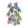
- PDB-9qfd: Cryo-EM structure of the fully cofilin-1-decorated actin filament... -
+ Open data

- Basic information
| Entry | Database: PDB / ID: 9qfd | |||||||||||||||
|---|---|---|---|---|---|---|---|---|---|---|---|---|---|---|---|---|
| Title | Cryo-EM structure of the fully cofilin-1-decorated actin filament (cofilactin) | |||||||||||||||
 Components Components |
| |||||||||||||||
 Keywords Keywords | STRUCTURAL PROTEIN / actin / cofilin / filament / cytoskeleton | |||||||||||||||
| Function / homology |  Function and homology information Function and homology informationcellular response to ether / cofilin-actin rod / positive regulation of protein localization to cell leading edge / positive regulation of establishment of cell polarity regulating cell shape / neural fold formation / negative regulation of unidimensional cell growth / positive regulation of barbed-end actin filament capping / negative regulation of lamellipodium assembly / negative regulation of postsynaptic density organization / actin filament fragmentation ...cellular response to ether / cofilin-actin rod / positive regulation of protein localization to cell leading edge / positive regulation of establishment of cell polarity regulating cell shape / neural fold formation / negative regulation of unidimensional cell growth / positive regulation of barbed-end actin filament capping / negative regulation of lamellipodium assembly / negative regulation of postsynaptic density organization / actin filament fragmentation / positive regulation of actin filament depolymerization / negative regulation of actin filament bundle assembly / modification of postsynaptic actin cytoskeleton / positive regulation of norepinephrine uptake / positive regulation of embryonic development / negative regulation of actin filament depolymerization / bBAF complex / cellular response to cytochalasin B / Formation of the embryonic stem cell BAF (esBAF) complex / npBAF complex / brahma complex / nBAF complex / Formation of the canonical BAF (cBAF) complex / regulation of transepithelial transport / host-mediated activation of viral process / actin filament severing / morphogenesis of a polarized epithelium / Formation of the polybromo-BAF (pBAF) complex / Formation of neuronal progenitor and neuronal BAF (npBAF and nBAF) / structural constituent of postsynaptic actin cytoskeleton / Formation of annular gap junctions / Formation of the dystrophin-glycoprotein complex (DGC) / positive regulation of synaptic plasticity / GBAF complex / regulation of dendritic spine morphogenesis / Gap junction degradation / Formation of the non-canonical BAF (ncBAF) complex / protein localization to adherens junction / regulation of G0 to G1 transition / establishment of spindle localization / Cell-extracellular matrix interactions / negative regulation of cell adhesion / dense body / Folding of actin by CCT/TriC / negative regulation of cell motility / Tat protein binding / actin filament depolymerization / RHO GTPases Activate ROCKs / negative regulation of cell size / postsynaptic actin cytoskeleton / RSC-type complex / cellular response to interleukin-6 / regulation of cell morphogenesis / Regulation of CDH1 Function / regulation of double-strand break repair / Prefoldin mediated transfer of substrate to CCT/TriC / regulation of nucleotide-excision repair / Adherens junctions interactions / adherens junction assembly / RHOF GTPase cycle / cell projection organization / apical protein localization / negative regulation of dendritic spine maintenance / Sensory processing of sound by outer hair cells of the cochlea / positive regulation of cell motility / regulation of mitotic metaphase/anaphase transition / tight junction / SWI/SNF complex / Sensory processing of sound by inner hair cells of the cochlea / Interaction between L1 and Ankyrins / neural crest cell migration / positive regulation of T cell differentiation / cortical actin cytoskeleton / apical junction complex / cellular response to insulin-like growth factor stimulus / phosphatidylinositol bisphosphate binding / positive regulation of double-strand break repair / establishment of cell polarity / maintenance of blood-brain barrier / regulation of norepinephrine uptake / positive regulation of dendritic spine development / transporter regulator activity / positive regulation of stem cell population maintenance / NuA4 histone acetyltransferase complex / Recycling pathway of L1 / Regulation of MITF-M-dependent genes involved in pigmentation / cortical cytoskeleton / positive regulation of proteolysis / establishment or maintenance of cell polarity / lamellipodium membrane / mitotic cytokinesis / nitric-oxide synthase binding / regulation of G1/S transition of mitotic cell cycle / brush border / Sema3A PAK dependent Axon repulsion / response to amino acid / EPH-ephrin mediated repulsion of cells / positive regulation of focal adhesion assembly / cellular response to interleukin-1 / negative regulation of cell differentiation Similarity search - Function | |||||||||||||||
| Biological species |  Homo sapiens (human) Homo sapiens (human) | |||||||||||||||
| Method | ELECTRON MICROSCOPY / helical reconstruction / cryo EM / Resolution: 2.61 Å | |||||||||||||||
 Authors Authors | Oosterheert, W. / Boiero Sanders, M. / Hofnagel, O. / Bieling, P. / Raunser, S. | |||||||||||||||
| Funding support |  Germany, European Union, 4items Germany, European Union, 4items
| |||||||||||||||
 Citation Citation | Journal: Cell / Year: 2025 Title: Choreography of rapid actin filament disassembly by coronin, cofilin, and AIP1. Authors: Wout Oosterheert / Micaela Boiero Sanders / Oliver Hofnagel / Peter Bieling / Stefan Raunser / Abstract: Rapid remodeling of actin filament (F-actin) networks is essential for the movement and morphogenesis of eukaryotic cells. The conserved actin-binding proteins coronin, cofilin, and actin-interacting ...Rapid remodeling of actin filament (F-actin) networks is essential for the movement and morphogenesis of eukaryotic cells. The conserved actin-binding proteins coronin, cofilin, and actin-interacting protein 1 (AIP1) act in synergy to promote rapid F-actin network disassembly, but the underlying mechanisms have remained elusive. Here, using cryo-electron microscopy (cryo-EM), we uncover the concerted molecular actions of coronin, cofilin, and AIP1 that lead to actin filament aging and severing. We find that the cooperative binding of coronin allosterically promotes inorganic phosphate release from F-actin and induces filament undertwisting, thereby priming the filament for cofilin binding. Cofilin then displaces coronin from the filament via a strand-restricted cooperative binding mechanism. The resulting cofilactin serves as a high-affinity platform for AIP1, which induces severing by acting as a clamp that disrupts inter-subunit filament contacts. In this "molecular squeezing" mechanism, AIP1 and not cofilin is responsible for filament severing. Our work redefines the role of key disassembly factors in actin dynamics. | |||||||||||||||
| History |
|
- Structure visualization
| Structure viewer | Molecule: MolmilJmol/JSmol |
|---|
- Downloads & links
-Download
| PDBx/mmCIF format | 9qfd.cif.gz | 867.5 KB | Display | PDBx/mmCIF format |
|---|---|---|---|---|
| PDB format | pdb9qfd.ent.gz | 577.6 KB | Display | PDB format |
| PDBx/mmJSON format | 9qfd.json.gz | Tree view | PDBx/mmJSON format | |
| Others |  Other downloads Other downloads |
-Validation report
| Arichive directory | https://data.pdbj.org/pub/pdb/validation_reports/qf/9qfdftp://data.pdbj.org/pub/pdb/validation_reports/qf/9qfd | HTTPS FTP |
|---|
-Related structure data
| Related structure data |  53106MC  9qewC  9qeyC  9qf2C  9qfbC  9qfeC  9qfgC  9qfjC  9qfkC  9qfoC  9qfqC  9qfwC C: citing same article ( M: map data used to model this data |
|---|---|
| Similar structure data |


-Links
 PDBj
PDBj


- Assembly
| Deposited unit | 
|
|---|---|
| 1 |
|
-Components
| #1: Protein | Mass: 41632.422 Da / Num. of mol.: 7 / Mutation: C272A Source method: isolated from a genetically manipulated source Details: actin filament / Source: (gene. exp.) Homo sapiens (human) / Gene: ACTB / Cell line (production host): BTI-Tnao38 / Production host:  Trichoplusia ni (cabbage looper) / References: UniProt: P60709 Trichoplusia ni (cabbage looper) / References: UniProt: P60709#2: Protein | Mass: 18532.531 Da / Num. of mol.: 7 Source method: isolated from a genetically manipulated source Details: Cofilin / Source: (gene. exp.) Homo sapiens (human) / Gene: CFL1, CFL / Production host:  #3: Chemical | ChemComp-ADP /   Mass: 427.201 Da / Num. of mol.: 7 / Source method: obtained synthetically / Formula: C10H15N5O10P2 / Feature type: SUBJECT OF INVESTIGATION / Comment: ADP, energy-carrying molecule*YM Mass: 427.201 Da / Num. of mol.: 7 / Source method: obtained synthetically / Formula: C10H15N5O10P2 / Feature type: SUBJECT OF INVESTIGATION / Comment: ADP, energy-carrying molecule*YM#4: Chemical | ChemComp-MG /   Mass: 24.305 Da / Num. of mol.: 7 / Source method: obtained synthetically / Formula: Mg / Feature type: SUBJECT OF INVESTIGATION Mass: 24.305 Da / Num. of mol.: 7 / Source method: obtained synthetically / Formula: Mg / Feature type: SUBJECT OF INVESTIGATIONHas ligand of interest | Y | Has protein modification | Y | |
|---|
-Experimental details
-Experiment
| Experiment | Method: ELECTRON MICROSCOPY |
|---|---|
| EM experiment | Aggregation state: FILAMENT / 3D reconstruction method: helical reconstruction |
- Sample preparation
| Component |
| |||||||||||||||||||||||||||||||||||
|---|---|---|---|---|---|---|---|---|---|---|---|---|---|---|---|---|---|---|---|---|---|---|---|---|---|---|---|---|---|---|---|---|---|---|---|---|
| Molecular weight |
| |||||||||||||||||||||||||||||||||||
| Source (natural) |
| |||||||||||||||||||||||||||||||||||
| Source (recombinant) |
| |||||||||||||||||||||||||||||||||||
| Buffer solution | pH: 7.1 Details: 1xKMEH (10 mM HEPES pH 7.1, 100 mM KCl, 2 mM MgCl2, 1 mM EGTA, 0.5 mM TCEP, 0.02% Tween20). | |||||||||||||||||||||||||||||||||||
| Buffer component |
| |||||||||||||||||||||||||||||||||||
| Specimen | Embedding applied: NO / Shadowing applied: NO / Staining applied: NO / Vitrification applied: YES | |||||||||||||||||||||||||||||||||||
| Specimen support | Grid material: COPPER / Grid mesh size: 200 divisions/in. / Grid type: Quantifoil R2/1 | |||||||||||||||||||||||||||||||||||
| Vitrification | Instrument: FEI VITROBOT MARK IV / Cryogen name: ETHANE-PROPANE / Humidity: 100 % / Chamber temperature: 286 K |
- Electron microscopy imaging
| Experimental equipment |  Model: Titan Krios / Image courtesy: FEI Company |
|---|---|
| Microscopy | Model: TFS KRIOS |
| Electron gun | Electron source:  FIELD EMISSION GUN / Accelerating voltage: 300 kV / Illumination mode: FLOOD BEAM FIELD EMISSION GUN / Accelerating voltage: 300 kV / Illumination mode: FLOOD BEAM |
| Electron lens | Mode: BRIGHT FIELD / Nominal magnification: 105000 X / Nominal defocus max: 2700 nm / Nominal defocus min: 1200 nm / Cs: 0.01 mm / C2 aperture diameter: 50 µm |
| Specimen holder | Cryogen: NITROGEN / Specimen holder model: FEI TITAN KRIOS AUTOGRID HOLDER |
| Image recording | Electron dose: 65.8 e/Å2 / Film or detector model: GATAN K3 BIOQUANTUM (6k x 4k) / Num. of grids imaged: 1 / Num. of real images: 14055 |
| EM imaging optics | Energyfilter name: GIF Bioquantum / Energyfilter slit width: 15 eV Spherical aberration corrector: The used Titan Krios G2 microscope contains an in-column Cs corrector. |
- Processing
| EM software |
| |||||||||||||||||||||||||||||||||||||||||||||||||||||||
|---|---|---|---|---|---|---|---|---|---|---|---|---|---|---|---|---|---|---|---|---|---|---|---|---|---|---|---|---|---|---|---|---|---|---|---|---|---|---|---|---|---|---|---|---|---|---|---|---|---|---|---|---|---|---|---|---|
| CTF correction | Details: Patch CTF / Type: PHASE FLIPPING AND AMPLITUDE CORRECTION | |||||||||||||||||||||||||||||||||||||||||||||||||||||||
| Helical symmerty | Angular rotation/subunit: -162.4 ° / Axial rise/subunit: 27.4 Å / Axial symmetry: C1 | |||||||||||||||||||||||||||||||||||||||||||||||||||||||
| Particle selection | Num. of particles selected: 10071093 | |||||||||||||||||||||||||||||||||||||||||||||||||||||||
| 3D reconstruction | Resolution: 2.61 Å / Resolution method: FSC 0.143 CUT-OFF / Num. of particles: 798636 / Symmetry type: HELICAL | |||||||||||||||||||||||||||||||||||||||||||||||||||||||
| Atomic model building | Protocol: FLEXIBLE FIT / Space: REAL / Details: Phenix real space refinement. | |||||||||||||||||||||||||||||||||||||||||||||||||||||||
| Atomic model building | PDB-ID: 6VAO Accession code: 6VAO / Source name: PDB / Type: experimental model | |||||||||||||||||||||||||||||||||||||||||||||||||||||||
| Refinement | Cross valid method: NONE Stereochemistry target values: GeoStd + Monomer Library + CDL v1.2 | |||||||||||||||||||||||||||||||||||||||||||||||||||||||
| Displacement parameters | Biso mean: 83.46 Å2 | |||||||||||||||||||||||||||||||||||||||||||||||||||||||
| Refine LS restraints |
|