 Movie
Movie Controller


+ Open data

- Basic information
| Entry | Database: PDB / ID: 9pa3 | |||||||||||||||||||||
|---|---|---|---|---|---|---|---|---|---|---|---|---|---|---|---|---|---|---|---|---|---|---|
| Title | 3-OH-PCP bound mu-opioid receptor in complex with Gi1 | |||||||||||||||||||||
 Components Components |
| |||||||||||||||||||||
 Keywords Keywords | SIGNALING PROTEIN / mu-opioid receptor / GPCR | |||||||||||||||||||||
| Function / homology |  Function and homology information Function and homology informationOpioid Signalling / beta-endorphin receptor activity / morphine receptor activity / negative regulation of Wnt protein secretion / G protein-coupled opioid receptor signaling pathway / negative regulation of nitric oxide biosynthetic process / adenylate cyclase-inhibiting G protein-coupled acetylcholine receptor signaling pathway / behavioral response to ethanol / positive regulation of neurogenesis / regulation of NMDA receptor activity ...Opioid Signalling / beta-endorphin receptor activity / morphine receptor activity / negative regulation of Wnt protein secretion / G protein-coupled opioid receptor signaling pathway / negative regulation of nitric oxide biosynthetic process / adenylate cyclase-inhibiting G protein-coupled acetylcholine receptor signaling pathway / behavioral response to ethanol / positive regulation of neurogenesis / regulation of NMDA receptor activity / neuropeptide binding / sensory perception / negative regulation of cytosolic calcium ion concentration / regulation of cellular response to stress / transmission of nerve impulse / G-protein alpha-subunit binding / G protein-coupled receptor signaling pathway, coupled to cyclic nucleotide second messenger / MECP2 regulates neuronal receptors and channels / voltage-gated calcium channel activity / sensory perception of pain / regulation of eating behavior / adenylate cyclase inhibitor activity / T cell migration / positive regulation of protein localization to cell cortex / positive regulation of relaxation of smooth muscle / Adenylate cyclase inhibitory pathway / D2 dopamine receptor binding / presynaptic modulation of chemical synaptic transmission / adenylate cyclase-inhibiting dopamine receptor signaling pathway / adenylate cyclase-inhibiting serotonin receptor signaling pathway / G protein-coupled serotonin receptor binding / regulation of G protein-coupled receptor signaling pathway / cellular response to forskolin / regulation of mitotic spindle organization / Peptide ligand-binding receptors / mast cell degranulation / chemokine-mediated signaling pathway / establishment of mitotic spindle orientation / neuropeptide signaling pathway / Regulation of insulin secretion / response to prostaglandin E / GABA-ergic synapse / positive regulation of cholesterol biosynthetic process / G protein-coupled receptor binding / response to peptide hormone / G protein-coupled receptor activity / G-protein beta/gamma-subunit complex binding / adenylate cyclase-inhibiting G protein-coupled receptor signaling pathway / adenylate cyclase-modulating G protein-coupled receptor signaling pathway / Olfactory Signaling Pathway / phospholipase C-activating G protein-coupled receptor signaling pathway / Activation of the phototransduction cascade / G protein-coupled acetylcholine receptor signaling pathway / G beta:gamma signalling through PLC beta / Presynaptic function of Kainate receptors / Thromboxane signalling through TP receptor / Activation of G protein gated Potassium channels / Inhibition of voltage gated Ca2+ channels via Gbeta/gamma subunits / G-protein activation / Glucagon signaling in metabolic regulation / G beta:gamma signalling through CDC42 / Prostacyclin signalling through prostacyclin receptor / GDP binding / Synthesis, secretion, and inactivation of Glucagon-like Peptide-1 (GLP-1) / photoreceptor disc membrane / G beta:gamma signalling through BTK / ADP signalling through P2Y purinoceptor 12 / Sensory perception of sweet, bitter, and umami (glutamate) taste / Glucagon-type ligand receptors / Adrenaline,noradrenaline inhibits insulin secretion / Vasopressin regulates renal water homeostasis via Aquaporins / G alpha (z) signalling events / Glucagon-like Peptide-1 (GLP1) regulates insulin secretion / ADP signalling through P2Y purinoceptor 1 / cellular response to catecholamine stimulus / G beta:gamma signalling through PI3Kgamma / ADORA2B mediated anti-inflammatory cytokines production / adenylate cyclase-activating dopamine receptor signaling pathway / cellular response to prostaglandin E stimulus / Cooperation of PDCL (PhLP1) and TRiC/CCT in G-protein beta folding / GPER1 signaling / presynapse / heterotrimeric G-protein complex / G alpha (12/13) signalling events / Inactivation, recovery and regulation of the phototransduction cascade / G-protein beta-subunit binding / extracellular vesicle / Thrombin signalling through proteinase activated receptors (PARs) / adenylate cyclase-activating G protein-coupled receptor signaling pathway / signaling receptor complex adaptor activity / spindle pole / ciliary basal body / Interleukin-4 and Interleukin-13 signaling / GTPase binding / midbody / G protein activity / cell cortex / Ca2+ pathway / High laminar flow shear stress activates signaling by PIEZO1 and PECAM1:CDH5:KDR in endothelial cells / G alpha (i) signalling events Similarity search - Function | |||||||||||||||||||||
| Biological species |  Homo sapiens (human) Homo sapiens (human)  Mus musculus (house mouse) Mus musculus (house mouse) | |||||||||||||||||||||
| Method | ELECTRON MICROSCOPY / single particle reconstruction / cryo EM / Resolution: 3.05 Å | |||||||||||||||||||||
 Authors Authors | Jiang, Q.R. / Han, J.M. / Fay, J.F. / Che, T. | |||||||||||||||||||||
| Funding support |  United States, 2items United States, 2items
| |||||||||||||||||||||
 Citation Citation | Journal: Nat Struct Mol Biol / Year: 2026 Title: Structural basis of opioid receptor activation by PCP and ketamine. Authors: Qianru Jiang / Jianming Han / Eve J Fine / Nokomis Ramos-Gonzalez / Vipin Ashok Rangari / Micaela V Ruiz / Madalyn L Critz / Carl-Mikael Suomivuori / Jing Wang / Talia L Albert / Kyle ...Authors: Qianru Jiang / Jianming Han / Eve J Fine / Nokomis Ramos-Gonzalez / Vipin Ashok Rangari / Micaela V Ruiz / Madalyn L Critz / Carl-Mikael Suomivuori / Jing Wang / Talia L Albert / Kyle Whiddon / Kunpeng Li / Michael J Robertson / Xi-Ping Huang / Benjamin B Land / Susruta Majumdar / Jonathan F Fay / Ron O Dror / Tao Che / Abstract: Ketamine offers rapid relief for treatment-resistant depression and severe pain in the clinic, providing immediate benefits that traditional medications often fail to deliver. While its antagonistic ...Ketamine offers rapid relief for treatment-resistant depression and severe pain in the clinic, providing immediate benefits that traditional medications often fail to deliver. While its antagonistic action at the N-methyl-D-aspartate receptor (NMDAR) is a key mechanism, ketamine's dual nature as both a promising treatment and a drug with abuse potential suggests its therapeutic effects extend beyond NMDAR inhibition. Here we provide structural evidence of human opioid receptors bound to ketamine and its parent analog phencyclidine (PCP), supporting that both ligands can directly bind and activate opioid receptors. The structures, together with site-directed mutagenesis and structure-activity relationship studies, identify key motifs involved in ketamine and PCP recognition and efficacy modulation. Furthermore, we determine the structure of the ligand-free state of human κ opioid receptor, revealing molecular details before ligand engagement. Compared to PCP, ketamine displays more notable binding dynamics in the orthosteric site that may contribute to its unique pharmacology at opioid receptors. Our findings highlight the importance of including opioid receptors to fully understand ketamine's versatility in clinical settings. | |||||||||||||||||||||
| History |
|
- Structure visualization
| Structure viewer | Molecule: MolmilJmol/JSmol |
|---|
- Downloads & links
-Download
| PDBx/mmCIF format | 9pa3.cif.gz | 236.4 KB | Display | PDBx/mmCIF format |
|---|---|---|---|---|
| PDB format | pdb9pa3.ent.gz | Display | PDB format | |
| PDBx/mmJSON format | 9pa3.json.gz | Tree view | PDBx/mmJSON format | |
| Others |  Other downloads Other downloads |
-Validation report
| Arichive directory | https://data.pdbj.org/pub/pdb/validation_reports/pa/9pa3ftp://data.pdbj.org/pub/pdb/validation_reports/pa/9pa3 | HTTPS FTP |
|---|
-Related structure data
| Related structure data |  71431MC  9pa1C  9pa2C  9pa4C  9pa5C  9pa6C M: map data used to model this data C: citing same article ( |
|---|---|
| Similar structure data |


-Links
 PDBj
PDBj























- Assembly
| Deposited unit | 
|
|---|---|
| 1 |
|
-Components
-Guanine nucleotide-binding protein ... , 3 types, 3 molecules BCA
| #1: Protein | Mass: 37342.785 Da / Num. of mol.: 1 Source method: isolated from a genetically manipulated source Source: (gene. exp.) Homo sapiens (human) / Gene: GNB1 / Production host:   Spodoptera frugiperda (fall armyworm) / References: UniProt: P62873 Spodoptera frugiperda (fall armyworm) / References: UniProt: P62873 |
|---|---|
| #2: Protein | Mass: 7861.143 Da / Num. of mol.: 1 Source method: isolated from a genetically manipulated source Source: (gene. exp.) Homo sapiens (human) / Gene: GNG2 / Production host: Spodoptera frugiperda (fall armyworm) / References: UniProt: P59768 |
| #3: Protein | Mass: 40414.047 Da / Num. of mol.: 1 Source method: isolated from a genetically manipulated source Source: (gene. exp.) Homo sapiens (human) / Gene: GNAI1 / Production host: Spodoptera frugiperda (fall armyworm) / References: UniProt: P63096 |
-Protein / Antibody / Non-polymers , 3 types, 3 molecules RE
| #4: Protein | Mass: 35105.863 Da / Num. of mol.: 1 Source method: isolated from a genetically manipulated source Source: (gene. exp.) Homo sapiens (human) / Gene: OPRM1, MOR1 / Production host: Spodoptera frugiperda (fall armyworm) / References: UniProt: P35372 |
|---|---|
| #5: Antibody | Mass: 26679.721 Da / Num. of mol.: 1 Source method: isolated from a genetically manipulated source Source: (gene. exp.) Mus musculus (house mouse) / Production host: Spodoptera frugiperda (fall armyworm) |
| #6: Chemical | ChemComp-A1CHE / Mass: 259.386 Da / Num. of mol.: 1 / Source method: obtained synthetically / Formula: C17H25NO / Feature type: SUBJECT OF INVESTIGATION |
-Details
| Has ligand of interest | Y |
|---|---|
| Has protein modification | Y |
-Experimental details
-Experiment
| Experiment | Method: ELECTRON MICROSCOPY |
|---|---|
| EM experiment | Aggregation state: PARTICLE / 3D reconstruction method: single particle reconstruction |
- Sample preparation
| Component | Name: The cryo-EM structure of MOR and 3-OH-PCP Ligand: 3-OH-PCP Type: COMPLEX / Entity ID: #1-#3, #5, #4 / Source: RECOMBINANT |
|---|---|
| Molecular weight | Experimental value: NO |
| Source (natural) | Organism: Homo sapiens (human) |
| Source (recombinant) | Organism: Spodoptera frugiperda (fall armyworm) |
| Buffer solution | pH: 7.4 |
| Specimen | Embedding applied: NO / Shadowing applied: NO / Staining applied: NO / Vitrification applied: YES |
| Vitrification | Instrument: FEI VITROBOT MARK IV / Cryogen name: ETHANE |
- Electron microscopy imaging
| Experimental equipment | 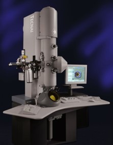 Model: Tecnai Polara / Image courtesy: FEI Company |
|---|---|
| Microscopy | Model: FEI POLARA 300 |
| Electron gun | Electron source:  FIELD EMISSION GUN / Accelerating voltage: 300 kV / Illumination mode: FLOOD BEAM FIELD EMISSION GUN / Accelerating voltage: 300 kV / Illumination mode: FLOOD BEAM |
| Electron lens | Mode: BRIGHT FIELD / Nominal defocus max: 2600 nm / Nominal defocus min: 200 nm |
| Image recording | Electron dose: 60 e/Å2 / Film or detector model: GATAN K3 (6k x 4k) |
- Processing
| EM software | Name: PHENIX / Version: 1.20.1_4487 / Category: model refinement | ||||||||||||||||||||||||
|---|---|---|---|---|---|---|---|---|---|---|---|---|---|---|---|---|---|---|---|---|---|---|---|---|---|
| CTF correction | Type: PHASE FLIPPING AND AMPLITUDE CORRECTION | ||||||||||||||||||||||||
| 3D reconstruction | Resolution: 3.05 Å / Resolution method: FSC 0.143 CUT-OFF / Num. of particles: 431685 / Symmetry type: POINT | ||||||||||||||||||||||||
| Refinement | Highest resolution: 3.05 Å Stereochemistry target values: REAL-SPACE (WEIGHTED MAP SUM AT ATOM CENTERS) | ||||||||||||||||||||||||
| Refine LS restraints |
|
