Movie
Movie Controller

[English] 日本語
 Yorodumi
- PDB-9ouk: DDB1-CRBN with Ikaros(ZF2) and DEG-47: composite map and model su... -
+ Open data

- Basic information
| Entry | Database: PDB / ID: 9ouk | |||||||||||||||
|---|---|---|---|---|---|---|---|---|---|---|---|---|---|---|---|---|
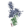
| Title | DDB1-CRBN with Ikaros(ZF2) and DEG-47: composite map and model submission | |||||||||||||||
 Components Components |
| |||||||||||||||
 Keywords Keywords | LIGASE / E3 ligases / Cullin RING Ligase / CRL4 / Cereblon / CRBN / molecular glues / IMiDs / Ikaros | |||||||||||||||
| Function / homology |  Function and homology information Function and homology informationlymphocyte differentiation / negative regulation of monoatomic ion transmembrane transport / positive regulation by virus of viral protein levels in host cell / spindle assembly involved in female meiosis / epigenetic programming in the zygotic pronuclei / NOTCH3 Intracellular Domain Regulates Transcription / UV-damage excision repair / biological process involved in interaction with symbiont / limb development / regulation of mitotic cell cycle phase transition ...lymphocyte differentiation / negative regulation of monoatomic ion transmembrane transport / positive regulation by virus of viral protein levels in host cell / spindle assembly involved in female meiosis / epigenetic programming in the zygotic pronuclei / NOTCH3 Intracellular Domain Regulates Transcription / UV-damage excision repair / biological process involved in interaction with symbiont / limb development / regulation of mitotic cell cycle phase transition / WD40-repeat domain binding / Cul4A-RING E3 ubiquitin ligase complex / Cul4-RING E3 ubiquitin ligase complex / Cul4B-RING E3 ubiquitin ligase complex / mesoderm development / ubiquitin ligase complex scaffold activity / negative regulation of reproductive process / negative regulation of developmental process / locomotory exploration behavior / viral release from host cell / cullin family protein binding / ectopic germ cell programmed cell death / pericentric heterochromatin / positive regulation of viral genome replication / positive regulation of Wnt signaling pathway / negative regulation of protein-containing complex assembly / proteasomal protein catabolic process / positive regulation of gluconeogenesis / erythrocyte differentiation / nucleotide-excision repair / sperm end piece / positive regulation of protein-containing complex assembly / regulation of circadian rhythm / Recognition of DNA damage by PCNA-containing replication complex / DNA Damage Recognition in GG-NER / Dual Incision in GG-NER / Wnt signaling pathway / Transcription-Coupled Nucleotide Excision Repair (TC-NER) / Formation of TC-NER Pre-Incision Complex / Formation of Incision Complex in GG-NER / Dual incision in TC-NER / positive regulation of protein catabolic process / Gap-filling DNA repair synthesis and ligation in TC-NER / cellular response to UV / rhythmic process / site of double-strand break / sperm principal piece / chromatin organization / Neddylation / sperm midpiece / Potential therapeutics for SARS / ubiquitin-dependent protein catabolic process / damaged DNA binding / transmembrane transporter binding / proteasome-mediated ubiquitin-dependent protein catabolic process / protein-macromolecule adaptor activity / chromosome, telomeric region / RNA polymerase II cis-regulatory region sequence-specific DNA binding / protein ubiquitination / DNA-binding transcription factor activity / protein domain specific binding / DNA repair / negative regulation of DNA-templated transcription / apoptotic process / DNA damage response / regulation of transcription by RNA polymerase II / negative regulation of apoptotic process / protein-containing complex binding / nucleolus / perinuclear region of cytoplasm / protein-containing complex / : / DNA binding / extracellular exosome / zinc ion binding / nucleoplasm / membrane / metal ion binding / identical protein binding / nucleus / cytoplasm / cytosol Similarity search - Function | |||||||||||||||
| Biological species |  Homo sapiens (human) Homo sapiens (human) | |||||||||||||||
| Method | ELECTRON MICROSCOPY / single particle reconstruction / cryo EM / Resolution: 2.69 Å | |||||||||||||||
 Authors Authors | Rizvi, Z. / Lander, G.C. | |||||||||||||||
| Funding support |  United States, 1items United States, 1items
| |||||||||||||||
 Citation Citation | Journal: Nature / Year: 2026 Title: Identification of an allosteric site on the E3 ligase adapter cereblon. Authors: Vanessa N Dippon / Zeba Rizvi / Anthony E Choudhry / Chun-Wa Chung / Ibrahim F Alkuraya / Wenqing Xu / Xavier B Tao / Anthony J Jurewicz / Jessica L Schneck / Wenqian Chen / Nicole M Curnutt ...Authors: Vanessa N Dippon / Zeba Rizvi / Anthony E Choudhry / Chun-Wa Chung / Ibrahim F Alkuraya / Wenqing Xu / Xavier B Tao / Anthony J Jurewicz / Jessica L Schneck / Wenqian Chen / Nicole M Curnutt / Farah Kabir / Kwok-Ho Chan / Markus A Queisser / Caterina Musetti / Han Dai / Gabriel C Lander / Andrew B Benowitz / Christina M Woo /  Abstract: Cereblon (CRBN) is the target of thalidomide derivatives that achieve therapeutic efficacy against some haematologic neoplasias by recruiting neosubstrates for degradation. Despite the intense ...Cereblon (CRBN) is the target of thalidomide derivatives that achieve therapeutic efficacy against some haematologic neoplasias by recruiting neosubstrates for degradation. Despite the intense investigation of orthosteric thalidomide derivatives, little is known about alternate binding sites on CRBN. Here we report an evolutionarily conserved cryptic allosteric binding site on CRBN. Small-molecule SB-405483 binds the allosteric site to cooperatively enhance the binding of orthosteric ligands and alter their neosubstrate degradation profiles. A survey of over 100 orthosteric ligands and their degradation targets reveals trends in the classes of compounds and neosubstrates in which degradation outcomes are enhanced or inhibited by SB-405483. Structural investigations provide a mechanistic basis for the effects of the allosteric ligand by shifting the conformational distribution of CRBN to a novel CRBN and increasing the CRBN state. The discovery of a cryptic allosteric binding site on CRBN that alters the functional effects of orthosteric ligands opens new directions with broad implications for improving the selectivity and efficacy of CRBN therapeutics. | |||||||||||||||
| History |
|
- Structure visualization
| Structure viewer | Molecule: MolmilJmol/JSmol |
|---|
- Downloads & links
-Download
| PDBx/mmCIF format | 9ouk.cif.gz | 259.2 KB | Display | PDBx/mmCIF format |
|---|---|---|---|---|
| PDB format | pdb9ouk.ent.gz | Display | PDB format | |
| PDBx/mmJSON format | 9ouk.json.gz | Tree view | PDBx/mmJSON format | |
| Others |  Other downloads Other downloads |
-Validation report
| Arichive directory | https://data.pdbj.org/pub/pdb/validation_reports/ou/9oukftp://data.pdbj.org/pub/pdb/validation_reports/ou/9ouk | HTTPS FTP |
|---|
-Related structure data
| Related structure data |  70867MC  9otyC  9oulC  9sfmC C: citing same article ( M: map data used to model this data |
|---|---|
| Similar structure data |


-Links
 PDBj
PDBj


- Assembly
| Deposited unit | 
|
|---|---|
| 1 |
|
-Components
| #1: Protein | Mass: 94389.234 Da / Num. of mol.: 1 Source method: isolated from a genetically manipulated source Details: The BPB domain is truncated (396-705 truncation with addition of a GNGNSG linker between BPA and BPC) from DDB1 and co-expressed with Cereblon (CRBN) in SF9 expression system, Source: (gene. exp.) Homo sapiens (human) / Gene: DDB1, XAP1 / Plasmid: pFASTBac / Cell line (production host): Sf9Production host:  Spodoptera aff. frugiperda 1 BOLD-2017 (butterflies/moths) Spodoptera aff. frugiperda 1 BOLD-2017 (butterflies/moths)References: UniProt: Q16531 | ||||||
|---|---|---|---|---|---|---|---|
| #2: Protein | Mass: 51563.758 Da / Num. of mol.: 1 Source method: isolated from a genetically manipulated source Source: (gene. exp.) Homo sapiens (human) / Gene: CRBN, AD-006 / Plasmid: pFASTBac / Cell line (production host): Sf9Production host: Spodoptera aff. frugiperda 1 BOLD-2017 (butterflies/moths)References: UniProt: Q96SW2 | ||||||
| #3: Protein | Mass: 6539.526 Da / Num. of mol.: 1 Source method: isolated from a genetically manipulated source Source: (gene. exp.) Homo sapiens (human) / Gene: IKZF1, IK1, IKAROS, LYF1, ZNFN1A1 / Plasmid: pet28A / Production host:  | ||||||
| #4: Chemical |   Mass: 65.409 Da / Num. of mol.: 2 / Source method: obtained synthetically / Formula: Zn Mass: 65.409 Da / Num. of mol.: 2 / Source method: obtained synthetically / Formula: Zn#5: Chemical | ChemComp-A1CEK / | Mass: 363.367 Da / Num. of mol.: 1 / Source method: obtained synthetically / Formula: C20H17N3O4 / Feature type: SUBJECT OF INVESTIGATION Has ligand of interest | Y | Has protein modification | N | |
-Experimental details
-Experiment
| Experiment | Method: ELECTRON MICROSCOPY |
|---|---|
| EM experiment | Aggregation state: PARTICLE / 3D reconstruction method: single particle reconstruction |
- Sample preparation
| Component | Name: DDB1(BPB deleted)-CRBN complex with ikaros ZF2-3 and DEG-47 was incubauted for 30 minutes and then plunge frozen over 1.2/1.3 UltrAufoil Type: COMPLEX / Entity ID: #1-#3 / Source: MULTIPLE SOURCES | ||||||||||||||||||||
|---|---|---|---|---|---|---|---|---|---|---|---|---|---|---|---|---|---|---|---|---|---|
| Molecular weight | Value: 0.15127264 MDa / Experimental value: YES | ||||||||||||||||||||
| Source (natural) | Organism: Homo sapiens (human) | ||||||||||||||||||||
| Source (recombinant) | Organism: Spodoptera aff. frugiperda 1 BOLD-2017 (butterflies/moths) Strain: sf9 / Cell: insect cell / Plasmid: pFASTBac | ||||||||||||||||||||
| Buffer solution | pH: 7 / Details: 10mM HEPES, 240mM NaCl, 3mM TCEP | ||||||||||||||||||||
| Buffer component |
| ||||||||||||||||||||
| Specimen | Conc.: 0.5 mg/ml / Embedding applied: NO / Shadowing applied: NO / Staining applied: NO / Vitrification applied: YES Details: The protein complex with ikaros ZF2-3 was incubated with IMiD DEG-47 and was incubated at 1:2:13 for 30 minutes and then frozen on 1.2/1.3 Au grids | ||||||||||||||||||||
| Specimen support | Grid material: GOLD / Grid mesh size: 300 divisions/in. / Grid type: UltrAuFoil R1.2/1.3 | ||||||||||||||||||||
| Vitrification | Instrument: HOMEMADE PLUNGER / Cryogen name: ETHANE / Humidity: 95 % / Chamber temperature: 277.15 K |
- Electron microscopy imaging
| Experimental equipment |  Model: Talos Arctica / Image courtesy: FEI Company |
|---|---|
| Microscopy | Model: FEI TALOS ARCTICA |
| Electron gun | Electron source:  FIELD EMISSION GUN / Accelerating voltage: 200 kV / Illumination mode: FLOOD BEAM FIELD EMISSION GUN / Accelerating voltage: 200 kV / Illumination mode: FLOOD BEAM |
| Electron lens | Mode: BRIGHT FIELD / Nominal magnification: 19000 X / Calibrated magnification: 189189 X / Nominal defocus max: 1200 nm / Nominal defocus min: 800 nm / Cs: 2.7 mm / C2 aperture diameter: 50 µm / Alignment procedure: COMA FREE |
| Specimen holder | Cryogen: NITROGEN / Specimen holder model: FEI TITAN KRIOS AUTOGRID HOLDER / Temperature (max): 77 K / Temperature (min): 70 K |
| Image recording | Average exposure time: 4.08 sec. / Electron dose: 50 e/Å2 / Film or detector model: TFS FALCON 4i (4k x 4k) / Num. of grids imaged: 1 / Num. of real images: 4915 |
| Image scans | Sampling size: 14 µm / Width: 4096 / Height: 4096 |
- Processing
| EM software |
| ||||||||||||||||||||||||||||||||||||||||
|---|---|---|---|---|---|---|---|---|---|---|---|---|---|---|---|---|---|---|---|---|---|---|---|---|---|---|---|---|---|---|---|---|---|---|---|---|---|---|---|---|---|
| CTF correction | Type: PHASE FLIPPING ONLY | ||||||||||||||||||||||||||||||||||||||||
| Particle selection | Num. of particles selected: 3488921 | ||||||||||||||||||||||||||||||||||||||||
| Symmetry | Point symmetry: C1 (asymmetric) | ||||||||||||||||||||||||||||||||||||||||
| 3D reconstruction | Resolution: 2.69 Å / Resolution method: FSC 0.143 CUT-OFF / Num. of particles: 412326 / Algorithm: FOURIER SPACE / Num. of class averages: 1 / Symmetry type: POINT | ||||||||||||||||||||||||||||||||||||||||
| Atomic model building | B value: 97.8 / Protocol: RIGID BODY FIT / Space: RECIPROCAL | ||||||||||||||||||||||||||||||||||||||||
| Atomic model building | 3D fitting-ID: 1 / Accession code: 8d7z / Initial refinement model-ID: 1 / PDB-ID: 8d7z / Source name: PDB / Type: experimental model
| ||||||||||||||||||||||||||||||||||||||||
| Refinement | Highest resolution: 2.69 Å Stereochemistry target values: REAL-SPACE (WEIGHTED MAP SUM AT ATOM CENTERS) | ||||||||||||||||||||||||||||||||||||||||
| Refine LS restraints |
|