[English] 日本語
 Yorodumi
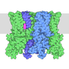
- PDB-9o93: Cryo-EM structure of KCa2.2_II/calmodulin channel in complex with... -
+ Open data
- Basic information
| Entry | Database: PDB / ID: 9o93 | |||||||||||||||||||||||||||||||||
|---|---|---|---|---|---|---|---|---|---|---|---|---|---|---|---|---|---|---|---|---|---|---|---|---|---|---|---|---|---|---|---|---|---|---|
| Title | Cryo-EM structure of KCa2.2_II/calmodulin channel in complex with rimtuzalcap | |||||||||||||||||||||||||||||||||
 Components Components |
| |||||||||||||||||||||||||||||||||
 Keywords Keywords | TRANSPORT PROTEIN / Ion channel / Small-conductance calcium-activated potassium channel | |||||||||||||||||||||||||||||||||
| Function / homology |  Function and homology information Function and homology informationCa2+ activated K+ channels / small conductance calcium-activated potassium channel activity / membrane repolarization during atrial cardiac muscle cell action potential / calcium-activated potassium channel activity / positive regulation of potassium ion transport / inward rectifier potassium channel activity / : / type 3 metabotropic glutamate receptor binding / establishment of protein localization to membrane / regulation of potassium ion transmembrane transport ...Ca2+ activated K+ channels / small conductance calcium-activated potassium channel activity / membrane repolarization during atrial cardiac muscle cell action potential / calcium-activated potassium channel activity / positive regulation of potassium ion transport / inward rectifier potassium channel activity / : / type 3 metabotropic glutamate receptor binding / establishment of protein localization to membrane / regulation of potassium ion transmembrane transport / negative regulation of ryanodine-sensitive calcium-release channel activity / organelle localization by membrane tethering / mitochondrion-endoplasmic reticulum membrane tethering / autophagosome membrane docking / negative regulation of calcium ion export across plasma membrane / regulation of cardiac muscle cell action potential / nitric-oxide synthase binding / presynaptic endocytosis / regulation of synaptic vesicle exocytosis / calcineurin-mediated signaling / alpha-actinin binding / smooth endoplasmic reticulum / adenylate cyclase binding / regulation of ryanodine-sensitive calcium-release channel activity / protein phosphatase activator activity / regulation of neuronal synaptic plasticity / regulation of synaptic vesicle endocytosis / detection of calcium ion / regulation of cardiac muscle contraction / postsynaptic cytosol / catalytic complex / phosphatidylinositol 3-kinase binding / calcium channel inhibitor activity / cellular response to interferon-beta / presynaptic cytosol / regulation of release of sequestered calcium ion into cytosol by sarcoplasmic reticulum / titin binding / regulation of calcium-mediated signaling / sperm midpiece / voltage-gated potassium channel complex / potassium ion transmembrane transport / T-tubule / calcium channel complex / regulation of heart rate / calyx of Held / response to amphetamine / nitric-oxide synthase regulator activity / adenylate cyclase activator activity / sarcomere / protein serine/threonine kinase activator activity / regulation of cytokinesis / spindle microtubule / positive regulation of receptor signaling pathway via JAK-STAT / calcium channel regulator activity / response to calcium ion / sarcolemma / modulation of chemical synaptic transmission / potassium ion transport / cellular response to type II interferon / Schaffer collateral - CA1 synapse / G2/M transition of mitotic cell cycle / Z disc / spindle pole / calcium-dependent protein binding / myelin sheath / growth cone / vesicle / dendritic spine / postsynaptic membrane / transmembrane transporter binding / calmodulin binding / protein domain specific binding / neuronal cell body / calcium ion binding / centrosome / protein kinase binding / glutamatergic synapse / cell surface / protein homodimerization activity / protein-containing complex / mitochondrion / nucleoplasm / membrane / plasma membrane / cytosol / cytoplasm Similarity search - Function | |||||||||||||||||||||||||||||||||
| Biological species |  | |||||||||||||||||||||||||||||||||
| Method | ELECTRON MICROSCOPY / single particle reconstruction / cryo EM / Resolution: 2.96 Å | |||||||||||||||||||||||||||||||||
 Authors Authors | Nam, Y.W. / Zhang, M. | |||||||||||||||||||||||||||||||||
| Funding support |  United States, 4items United States, 4items
| |||||||||||||||||||||||||||||||||
 Citation Citation |  Journal: Nat Commun / Year: 2026 Journal: Nat Commun / Year: 2026Title: Structural basis for the subtype-selectivity of K2.2 channel activators. Authors: Young-Woo Nam / Alena Ramanishka / Yang Xu / Rose Marie Haynes Yasuda / Joshua A Nasburg / Dohyun Im / Meng Cui / K George Chandy / Heike Wulff / Miao Zhang /    Abstract: Small-conductance (K2.2) and intermediate-conductance (K3.1) Ca-activated K channels are gated by a Ca-calmodulin dependent mechanism. NS309 potentiates the activity of both K2.2 and K3.1, while ...Small-conductance (K2.2) and intermediate-conductance (K3.1) Ca-activated K channels are gated by a Ca-calmodulin dependent mechanism. NS309 potentiates the activity of both K2.2 and K3.1, while rimtuzalcap selectively activates K2.2. Rimtuzalcap has been used in clinical trials for the treatment of spinocerebellar ataxia and essential tremor. We report cryo-electron microscopy structures of NS309-bound K2.2 and K3.1, in addition to structures of rimtuzalcap-bound K2.2 and mutant K3.1_R355K. The different conformations of calmodulin and the cytoplasmic HC helices in the two channels underlie the subtype-selectivity of rimtuzalcap for K2.2. NS309 binds to pre-existing pockets in both channels, while the bulkier rimtuzalcap binds in an induced-fit pocket in K2.2 requiring conformational changes. In K2.2, calmodulin's N-lobes are sufficiently far apart to enable conformational changes to accommodate either NS309 or rimtuzalcap. In K3.1, calmodulin's N-lobes are closer to each other and constrained by K3.1's HC helices, which allows binding of NS309 but not rimtuzalcap. Replacement of arginine-355 in K3.1's HB helix with lysine (K3.1_R355K) allows the binding of rimtuzalcap and renders the mutant channel sensitive to rimtuzalcap. These structures provide a framework for structure-based drug design targeting K2.2 channels. #1:  Journal: Res Sq / Year: 2025 Journal: Res Sq / Year: 2025Title: Structural basis for the subtype-selectivity of K2.2 channel activators. Authors: Miao Zhang / Young-Woo Nam / Alena Ramanishka / Yang Xu / Rose Marie Yasuda / Dohyun Im / Meng Cui / George Chandy / Heike Wulff /  Abstract: Small-conductance (K2.2) and intermediate-conductance (K3.1) Ca-activated K channels are gated by a Ca-calmodulin dependent mechanism. NS309 potentiates the activity of both K2.2 and K3.1, while ...Small-conductance (K2.2) and intermediate-conductance (K3.1) Ca-activated K channels are gated by a Ca-calmodulin dependent mechanism. NS309 potentiates the activity of both K2.2 and K3.1, while rimtuzalcap selectively activates K2.2. Rimtuzalcap has been used in clinical trials for the treatment of spinocerebellar ataxia and essential tremor. We report cryo-electron microscopy structures of K2.2 channels bound with NS309 and rimtuzalcap, in addition to K3.1 channels with NS309. The different conformations of calmodulin and the cytoplasmic HC helices in the two channels underlie the subtype-selectivity of rimtuzalcap for K2.2. Calmodulin's N-lobes in the K2.2 structure are far apart and undergo conformational changes to accommodate either NS309 or rimtuzalcap. Calmodulin's Nlobes in the K3.1 structure are closer to each other and are constrained by the HC helices of K3.1, which allows binding of NS309 but not of the bulkier rimtuzalcap. These structures provide a framework for structure-based drug design targeting K2.2 channels. | |||||||||||||||||||||||||||||||||
| History |
|
- Structure visualization
| Structure viewer | Molecule:  Molmil Molmil Jmol/JSmol Jmol/JSmol |
|---|
- Downloads & links
- Download
| PDBx/mmCIF format |  9o93.cif.gz 9o93.cif.gz | 402.8 KB | Display |  PDBx/mmCIF format PDBx/mmCIF format |
|---|---|---|---|---|
| PDB format |  pdb9o93.ent.gz pdb9o93.ent.gz | Display |  PDB format PDB format | |
| PDBx/mmJSON format |  9o93.json.gz 9o93.json.gz | Tree view |  PDBx/mmJSON format PDBx/mmJSON format | |
| Others |  Other downloads Other downloads |
-Validation report
| Summary document |  9o93_validation.pdf.gz 9o93_validation.pdf.gz | 1.5 MB | Display |  wwPDB validaton report wwPDB validaton report |
|---|---|---|---|---|
| Full document |  9o93_full_validation.pdf.gz 9o93_full_validation.pdf.gz | 1.5 MB | Display | |
| Data in XML |  9o93_validation.xml.gz 9o93_validation.xml.gz | 63.2 KB | Display | |
| Data in CIF |  9o93_validation.cif.gz 9o93_validation.cif.gz | 96.7 KB | Display | |
| Arichive directory |  https://data.pdbj.org/pub/pdb/validation_reports/o9/9o93 https://data.pdbj.org/pub/pdb/validation_reports/o9/9o93 ftp://data.pdbj.org/pub/pdb/validation_reports/o9/9o93 ftp://data.pdbj.org/pub/pdb/validation_reports/o9/9o93 | HTTPS FTP |
-Related structure data
| Related structure data |  70240MC  9o85C  9oa8C  9y5qC  9ydzC M: map data used to model this data C: citing same article ( |
|---|---|
| Similar structure data | Similarity search - Function & homology  F&H Search F&H Search |
- Links
Links
- Assembly
| Deposited unit | 
|
|---|---|
| 1 |
|
- Components
| #1: Protein | Mass: 43349.289 Da / Num. of mol.: 4 Source method: isolated from a genetically manipulated source Source: (gene. exp.)   Homo sapiens (human) / References: UniProt: P70604 Homo sapiens (human) / References: UniProt: P70604#2: Protein | Mass: 16277.873 Da / Num. of mol.: 4 Source method: isolated from a genetically manipulated source Source: (gene. exp.)   Homo sapiens (human) / References: UniProt: P0DP29 Homo sapiens (human) / References: UniProt: P0DP29#3: Chemical | #4: Chemical | ChemComp-A1B92 / Mass: 378.420 Da / Num. of mol.: 4 / Source method: obtained synthetically / Formula: C18H24F2N6O / Feature type: SUBJECT OF INVESTIGATION #5: Water | ChemComp-HOH / | Has ligand of interest | Y | Has protein modification | N | |
|---|
-Experimental details
-Experiment
| Experiment | Method: ELECTRON MICROSCOPY |
|---|---|
| EM experiment | Aggregation state: PARTICLE / 3D reconstruction method: single particle reconstruction |
- Sample preparation
| Component | Name: RatKCa2.2_II/calmodulin channel in complex with rimtuzalcap Type: COMPLEX / Entity ID: #1-#2 / Source: RECOMBINANT |
|---|---|
| Molecular weight | Value: 0.22973 MDa / Experimental value: NO |
| Source (natural) | Organism:  Homo sapiens (human) Homo sapiens (human) |
| Source (recombinant) | Organism:  Homo sapiens (human) / Cell: HEK293 / Plasmid: pGEBacMam Homo sapiens (human) / Cell: HEK293 / Plasmid: pGEBacMam |
| Buffer solution | pH: 8 |
| Specimen | Conc.: 2 mg/ml / Embedding applied: NO / Shadowing applied: NO / Staining applied: NO / Vitrification applied: YES |
| Vitrification | Cryogen name: ETHANE |
- Electron microscopy imaging
| Experimental equipment |  Model: Titan Krios / Image courtesy: FEI Company |
|---|---|
| Microscopy | Model: TFS KRIOS |
| Electron gun | Electron source:  FIELD EMISSION GUN / Accelerating voltage: 300 kV / Illumination mode: FLOOD BEAM FIELD EMISSION GUN / Accelerating voltage: 300 kV / Illumination mode: FLOOD BEAM |
| Electron lens | Mode: BRIGHT FIELD / Nominal defocus max: 2300 nm / Nominal defocus min: 1300 nm |
| Specimen holder | Specimen holder model: FEI TITAN KRIOS AUTOGRID HOLDER |
| Image recording | Electron dose: 50 e/Å2 / Film or detector model: FEI FALCON IV (4k x 4k) |
- Processing
| EM software |
| ||||||||||||||||||||||||
|---|---|---|---|---|---|---|---|---|---|---|---|---|---|---|---|---|---|---|---|---|---|---|---|---|---|
| CTF correction | Type: PHASE FLIPPING AND AMPLITUDE CORRECTION | ||||||||||||||||||||||||
| 3D reconstruction | Resolution: 2.96 Å / Resolution method: FSC 0.143 CUT-OFF / Num. of particles: 102190 / Symmetry type: POINT | ||||||||||||||||||||||||
| Refinement | Highest resolution: 2.96 Å Stereochemistry target values: REAL-SPACE (WEIGHTED MAP SUM AT ATOM CENTERS) | ||||||||||||||||||||||||
| Refine LS restraints |
|
 Movie
Movie Controller


 PDBj
PDBj


