 Movie
Movie Controller

[English] 日本語
 Yorodumi
- PDB-9o11: Kv2.1 with voltage sensor in the up conformation under low potassium -
+ Open data

- Basic information
| Entry | Database: PDB / ID: 9o11 | ||||||
|---|---|---|---|---|---|---|---|
| Title | Kv2.1 with voltage sensor in the up conformation under low potassium | ||||||
 Components Components | Potassium voltage-gated channel subfamily B member 1 | ||||||
 Keywords Keywords | MEMBRANE PROTEIN / voltage-gated potassium channel | ||||||
| Function / homology |  Function and homology information Function and homology informationregulation of action potential / regulation of motor neuron apoptotic process / clustering of voltage-gated potassium channels / positive regulation of long-term synaptic depression / positive regulation of catecholamine secretion / positive regulation of norepinephrine secretion / cholinergic synapse / potassium ion export across plasma membrane / positive regulation of calcium ion-dependent exocytosis / delayed rectifier potassium channel activity ...regulation of action potential / regulation of motor neuron apoptotic process / clustering of voltage-gated potassium channels / positive regulation of long-term synaptic depression / positive regulation of catecholamine secretion / positive regulation of norepinephrine secretion / cholinergic synapse / potassium ion export across plasma membrane / positive regulation of calcium ion-dependent exocytosis / delayed rectifier potassium channel activity / proximal dendrite / : / Voltage gated Potassium channels / outward rectifier potassium channel activity / response to L-glutamate / postsynaptic specialization membrane / glutamate receptor signaling pathway / neuronal cell body membrane / lateral plasma membrane / action potential / potassium channel regulator activity / positive regulation of protein targeting to membrane / voltage-gated potassium channel activity / response to axon injury / voltage-gated potassium channel complex / potassium ion transmembrane transport / cellular response to nutrient levels / cellular response to calcium ion / dendrite membrane / protein localization to plasma membrane / SNARE binding / cellular response to glucose stimulus / protein homooligomerization / sarcolemma / negative regulation of insulin secretion / Glucagon-like Peptide-1 (GLP1) regulates insulin secretion / glucose homeostasis / transmembrane transporter binding / perikaryon / postsynaptic membrane / apical plasma membrane / protein heterodimerization activity / axon / dendrite / perinuclear region of cytoplasm / cell surface / plasma membrane Similarity search - Function | ||||||
| Biological species |  Homo sapiens (human) Homo sapiens (human) | ||||||
| Method | ELECTRON MICROSCOPY / single particle reconstruction / cryo EM / Resolution: 3.3 Å | ||||||
 Authors Authors | Mandala, V.S. / MacKinnon, R. | ||||||
| Funding support |  United States, 1items United States, 1items
| ||||||
 Citation Citation | Journal: Proc Natl Acad Sci U S A / Year: 2025 Title: Electric field-induced pore constriction in the human K2.1 channel. Authors: Venkata Shiva Mandala / Roderick MacKinnon / Abstract: Gating in voltage-dependent ion channels is regulated by the transmembrane voltage. This form of regulation is enabled by voltage-sensing domains (VSDs) that respond to transmembrane voltage ...Gating in voltage-dependent ion channels is regulated by the transmembrane voltage. This form of regulation is enabled by voltage-sensing domains (VSDs) that respond to transmembrane voltage differences by changing their conformation and exerting force on the pore to open or close it. Here, we use cryogenic electron microscopy to study the neuronal K2.1 channel in lipid vesicles with and without a voltage difference across the membrane. Hyperpolarizing voltage differences displace the positively charged S4 helix in the voltage sensor by one helical turn (~5 Å). When this displacement occurs, the S4 helix changes its contact with the pore at two different interfaces. When these changes are observed in fewer than four voltage sensors, the pore remains open, but when they are observed in all four voltage sensors, the pore constricts. The constriction occurs because the S4 helix, as it displaces inward, squeezes the right-handed helical bundle of pore-lining S6 helices. A similar conformational change occurs upon hyperpolarization of the EAG1 channel but with two helical turns displaced instead of one. Therefore, while K2.1 and EAG1 are from distinct architectural classes of voltage-dependent ion channels, called domain-swapped and non-domain-swapped, the way the voltage sensors gate their pores is very similar. | ||||||
| History |
|
- Structure visualization
| Structure viewer | Molecule: MolmilJmol/JSmol |
|---|
- Downloads & links
-Download
| PDBx/mmCIF format | 9o11.cif.gz | 441.2 KB | Display | PDBx/mmCIF format |
|---|---|---|---|---|
| PDB format | pdb9o11.ent.gz | 345.6 KB | Display | PDB format |
| PDBx/mmJSON format | 9o11.json.gz | Tree view | PDBx/mmJSON format | |
| Others |  Other downloads Other downloads |
-Validation report
| Arichive directory | https://data.pdbj.org/pub/pdb/validation_reports/o1/9o11ftp://data.pdbj.org/pub/pdb/validation_reports/o1/9o11 | HTTPS FTP |
|---|
-Related structure data
| Related structure data |  49994MC  9o10C 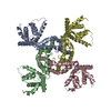 9o12C  9o13C M: map data used to model this data C: citing same article ( |
|---|---|
| Similar structure data |


-Links
 PDBj
PDBj


- Assembly
| Deposited unit | 
|
|---|---|
| 1 |
|
-Components
| #1: Protein | Mass: 96001.711 Da / Num. of mol.: 4 Source method: isolated from a genetically manipulated source Source: (gene. exp.) Homo sapiens (human) / Gene: KCNB1 / Production host: Homo sapiens (human) / References: UniProt: Q14721Has protein modification | N | |
|---|
-Experimental details
-Experiment
| Experiment | Method: ELECTRON MICROSCOPY |
|---|---|
| EM experiment | Aggregation state: PARTICLE / 3D reconstruction method: single particle reconstruction |
- Sample preparation
| Component | Name: human Kv2.1 / Type: CELL / Entity ID: all / Source: RECOMBINANT |
|---|---|
| Molecular weight | Experimental value: NO |
| Source (natural) | Organism: Homo sapiens (human) |
| Source (recombinant) | Organism: Homo sapiens (human) |
| Buffer solution | pH: 8 |
| Specimen | Conc.: 0.2 mg/ml / Embedding applied: NO / Shadowing applied: NO / Staining applied: NO / Vitrification applied: YES |
| Vitrification | Instrument: FEI VITROBOT MARK IV / Cryogen name: ETHANE / Humidity: 100 % / Chamber temperature: 293 K |
- Electron microscopy imaging
| Experimental equipment |  Model: Titan Krios / Image courtesy: FEI Company |
|---|---|
| Microscopy | Model: TFS KRIOS |
| Electron gun | Electron source:  FIELD EMISSION GUN / Accelerating voltage: 300 kV / Illumination mode: FLOOD BEAM FIELD EMISSION GUN / Accelerating voltage: 300 kV / Illumination mode: FLOOD BEAM |
| Electron lens | Mode: BRIGHT FIELD / Nominal defocus max: 2000 nm / Nominal defocus min: 1000 nm |
| Image recording | Electron dose: 60 e/Å2 / Film or detector model: FEI FALCON IV (4k x 4k) |
- Processing
| EM software | Name: PHENIX / Category: model refinement | ||||||||||||||||||||||||
|---|---|---|---|---|---|---|---|---|---|---|---|---|---|---|---|---|---|---|---|---|---|---|---|---|---|
| CTF correction | Type: PHASE FLIPPING AND AMPLITUDE CORRECTION | ||||||||||||||||||||||||
| Symmetry | Point symmetry: C4 (4 fold cyclic) | ||||||||||||||||||||||||
| 3D reconstruction | Resolution: 3.3 Å / Resolution method: FSC 0.143 CUT-OFF / Num. of particles: 47718 / Symmetry type: POINT | ||||||||||||||||||||||||
| Refinement | Highest resolution: 3.3 Å Stereochemistry target values: REAL-SPACE (WEIGHTED MAP SUM AT ATOM CENTERS) | ||||||||||||||||||||||||
| Refine LS restraints |
|
