[English] 日本語
Yorodumi
- EMDB-49996: Kv2.1 with 4 voltage sensors down and a constricted pore -

+
Open data


ID or keywords:

Loading...

-
Basic information

Entry
Database: EMDB / ID: EMD-49996
TitleKv2.1 with 4 voltage sensors down and a constricted pore
Map data
Sample
  • Cell: human Kv2.1
    • Protein or peptide: Potassium voltage-gated channel subfamily B member 1
Keywordsvoltage-gated potassium channel / MEMBRANE PROTEIN
Function / homology
Function and homology information


regulation of action potential / positive regulation of long-term synaptic depression / regulation of motor neuron apoptotic process / clustering of voltage-gated potassium channels / positive regulation of norepinephrine secretion / positive regulation of catecholamine secretion / positive regulation of calcium ion-dependent exocytosis / potassium ion export across plasma membrane / cholinergic synapse / proximal dendrite ...regulation of action potential / positive regulation of long-term synaptic depression / regulation of motor neuron apoptotic process / clustering of voltage-gated potassium channels / positive regulation of norepinephrine secretion / positive regulation of catecholamine secretion / positive regulation of calcium ion-dependent exocytosis / potassium ion export across plasma membrane / cholinergic synapse / proximal dendrite / delayed rectifier potassium channel activity / Voltage gated Potassium channels / outward rectifier potassium channel activity / vesicle docking involved in exocytosis / postsynaptic specialization membrane / glutamate receptor signaling pathway / neuronal cell body membrane / response to L-glutamate / action potential / positive regulation of protein targeting to membrane / response to axon injury / lateral plasma membrane / cellular response to nutrient levels / negative regulation of insulin secretion / dendrite membrane / voltage-gated potassium channel complex / potassium ion transmembrane transport / cellular response to calcium ion / SNARE binding / protein localization to plasma membrane / cellular response to glucose stimulus / sarcolemma / protein homooligomerization / Glucagon-like Peptide-1 (GLP1) regulates insulin secretion / glucose homeostasis / perikaryon / postsynaptic membrane / transmembrane transporter binding / apical plasma membrane / protein heterodimerization activity / axon / dendrite / perinuclear region of cytoplasm / cell surface / plasma membrane
Similarity search - Function
Potassium channel, voltage dependent, Kv2.1 / Potassium channel, voltage dependent, Kv2 / Kv2 voltage-gated K+ channel / Potassium channel, voltage dependent, Kv / Potassium channel tetramerisation-type BTB domain / BTB/POZ domain / Voltage-gated potassium channel / Broad-Complex, Tramtrack and Bric a brac / Voltage-dependent channel domain superfamily / BTB/POZ domain ...Potassium channel, voltage dependent, Kv2.1 / Potassium channel, voltage dependent, Kv2 / Kv2 voltage-gated K+ channel / Potassium channel, voltage dependent, Kv / Potassium channel tetramerisation-type BTB domain / BTB/POZ domain / Voltage-gated potassium channel / Broad-Complex, Tramtrack and Bric a brac / Voltage-dependent channel domain superfamily / BTB/POZ domain / SKP1/BTB/POZ domain superfamily / Ion transport domain / Ion transport protein
Similarity search - Domain/homology
Potassium voltage-gated channel subfamily B member 1
Similarity search - Component
Biological speciesHomo sapiens (human)
Methodsingle particle reconstruction / cryo EM / Resolution: 5.8 Å
AuthorsMandala VS / MacKinnon R
Funding support United States, 1 items
OrganizationGrant numberCountry
Howard Hughes Medical Institute (HHMI) United States
CitationJournal: Proc Natl Acad Sci U S A / Year: 2025
Title: Electric field-induced pore constriction in the human K2.1 channel.
Authors: Venkata Shiva Mandala / Roderick MacKinnon /
Abstract: Gating in voltage-dependent ion channels is regulated by the transmembrane voltage. This form of regulation is enabled by voltage-sensing domains (VSDs) that respond to transmembrane voltage ...Gating in voltage-dependent ion channels is regulated by the transmembrane voltage. This form of regulation is enabled by voltage-sensing domains (VSDs) that respond to transmembrane voltage differences by changing their conformation and exerting force on the pore to open or close it. Here, we use cryogenic electron microscopy to study the neuronal K2.1 channel in lipid vesicles with and without a voltage difference across the membrane. Hyperpolarizing voltage differences displace the positively charged S4 helix in the voltage sensor by one helical turn (~5 Å). When this displacement occurs, the S4 helix changes its contact with the pore at two different interfaces. When these changes are observed in fewer than four voltage sensors, the pore remains open, but when they are observed in all four voltage sensors, the pore constricts. The constriction occurs because the S4 helix, as it displaces inward, squeezes the right-handed helical bundle of pore-lining S6 helices. A similar conformational change occurs upon hyperpolarization of the EAG1 channel but with two helical turns displaced instead of one. Therefore, while K2.1 and EAG1 are from distinct architectural classes of voltage-dependent ion channels, called domain-swapped and non-domain-swapped, the way the voltage sensors gate their pores is very similar.
History
DepositionApr 3, 2025-
Header (metadata) releaseMay 28, 2025-
Map releaseMay 28, 2025-
UpdateMay 28, 2025-
Current statusMay 28, 2025Processing site: RCSB / Status: Released

-
Structure visualization
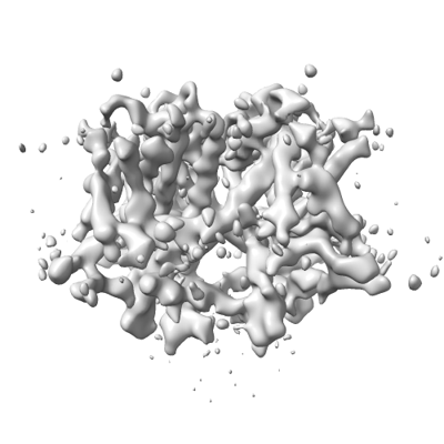
Supplemental images

Downloads & links

-
Map

FileDownload / File: emd_49996.map.gz / Format: CCP4 / Size: 91.1 MB / Type: IMAGE STORED AS FLOATING POINT NUMBER (4 BYTES)
Projections & slices

Image control

Size
Brightness
Contrast
Others
AxesZ (Sec.)Y (Row.)X (Col.)
0.74 Å/pix.
x 288 pix.
= 213.984 Å
0.74 Å/pix.
x 288 pix.
= 213.984 Å
0.74 Å/pix.
x 288 pix.
= 213.984 Å

Surface

Projections

Slices (1/3)

Slices (1/2)

Slices (2/3)

Images are generated by Spider.

Voxel sizeX=Y=Z: 0.743 Å
Density
Contour LevelBy AUTHOR: 0.0017
Minimum - Maximum-0.003127396 - 0.0055722487
Average (Standard dev.)0.000045332225 (±0.000501575)
SymmetrySpace group: 1
Details

EMDB XML:

Map geometry
Axis orderXYZ
Origin000
Dimensions288288288
Spacing288288288
CellA=B=C: 213.984 Å
α=β=γ: 90.0 °

-
Supplemental data

-
Additional map: sharpened map

Fileemd_49996_additional_1.map
Annotationsharpened map
Projections & Slices
AxesZYX

Projections

Slices (1/2)
Density Histograms

-
Half map: #2

Fileemd_49996_half_map_1.map
Projections & Slices
AxesZYX

Projections

Slices (1/2)
Density Histograms

-
Half map: #1

Fileemd_49996_half_map_2.map
Projections & Slices
AxesZYX

Projections

Slices (1/2)
Density Histograms

-
Sample components

-
Entire : human Kv2.1

EntireName: human Kv2.1
Components
  • Cell: human Kv2.1
    • Protein or peptide: Potassium voltage-gated channel subfamily B member 1

-
Supramolecule #1: human Kv2.1

SupramoleculeName: human Kv2.1 / type: cell / ID: 1 / Parent: 0 / Macromolecule list: all
Source (natural)Organism: Homo sapiens (human)

-
Macromolecule #1: Potassium voltage-gated channel subfamily B member 1

MacromoleculeName: Potassium voltage-gated channel subfamily B member 1 / type: protein_or_peptide / ID: 1 / Number of copies: 4 / Enantiomer: LEVO
Source (natural)Organism: Homo sapiens (human)
Molecular weightTheoretical: 96.001711 KDa
Recombinant expressionOrganism: Homo sapiens (human)
SequenceString: MPAGMTKHGS RSTSSLPPEP MEIVRSKACS RRVRLNVGGL AHEVLWRTLD RLPRTRLGKL RDCNTHDSLL EVCDDYSLDD NEYFFDRHP GAFTSILNFY RTGRLHMMEE MCALSFSQEL DYWGIDEIYL ESCCQARYHQ KKEQMNEELK REAETLRERE G EEFDNTCC ...String:
MPAGMTKHGS RSTSSLPPEP MEIVRSKACS RRVRLNVGGL AHEVLWRTLD RLPRTRLGKL RDCNTHDSLL EVCDDYSLDD NEYFFDRHP GAFTSILNFY RTGRLHMMEE MCALSFSQEL DYWGIDEIYL ESCCQARYHQ KKEQMNEELK REAETLRERE G EEFDNTCC AEKRKKLWDL LEKPNSSVAA KILAIISIMF IVLSTIALSL NTLPELQSLD EFGQSTDNPQ LAHVEAVCIA WF TMEYLLR FLSSPKKWKF FKGPLNAIDL LAILPYYVTI FLTESNKSVL QFQNVRRVVQ IFRIMRILRI LKLARHSTGL QSL GFTLRR SYNELGLLIL FLAMGIMIFS SLVFFAEKDE DDTKFKSIPA SFWWATITMT TVGYGDIYPK TLLGKIVGGL CCIA GVLVI ALPIPIIVNN FSEFYKEQKR QEKAIKRREA LERAKRNGSI VSMNMKDAFA RSIEMMDIVV EKNGENMGKK DKVQD NHLS PNKWKWTKRT LSETSSSKSF ETKEQGSPEK ARSSSSPQHL NVQQLEDMYN KMAKTQSQPI LNTKESAAQS KPKEEL EME SIPSPVAPLP TRTEGVIDMR SMSSIDSFIS CATDFPEATR FSHSPLTSLP SKTGGSTAPE VGWRGALGAS GGRFVEA NP SPDASQHSSF FIESPKSSMK TNNPLKLRAL KVNFMEGDPS PLLPVLGMYH DPLRNRGSAA AAVAGLECAT LLDKAVLS P ESSIYTTASA KTPPRSPEKH TAIAFNFEAG VHQYIDADTD DEGQLLYSVD SSPPKSLPGS TSPKFSTGTR SEKNHFESS PLPTSPKFLR QNCIYSTEAL TGKGPSGQEK CKLENHISPD VRVLPGGGAH GSTRDQSI

UniProtKB: Potassium voltage-gated channel subfamily B member 1

-
Experimental details

-
Structure determination

Methodcryo EM
Processingsingle particle reconstruction
Aggregation stateparticle

-
Sample preparation

Concentration0.2 mg/mL
BufferpH: 8
VitrificationCryogen name: ETHANE / Chamber humidity: 100 % / Chamber temperature: 293 K / Instrument: FEI VITROBOT MARK IV

-
Electron microscopy

MicroscopeTFS KRIOS
Image recordingFilm or detector model: FEI FALCON IV (4k x 4k) / Average electron dose: 60.0 e/Å2
Electron beamAcceleration voltage: 300 kV / Electron source: FIELD EMISSION GUN
Electron opticsIllumination mode: FLOOD BEAM / Imaging mode: BRIGHT FIELD / Nominal defocus max: 2.0 µm / Nominal defocus min: 1.0 µm
Experimental equipment
Model: Titan Krios / Image courtesy: FEI Company

+
Image processing

CTF correctionType: PHASE FLIPPING AND AMPLITUDE CORRECTION
Startup modelType of model: INSILICO MODEL
Final reconstructionApplied symmetry - Point group: C4 (4 fold cyclic) / Resolution.type: BY AUTHOR / Resolution: 5.8 Å / Resolution method: FSC 0.143 CUT-OFF / Number images used: 2251
Initial angle assignmentType: MAXIMUM LIKELIHOOD
Final angle assignmentType: MAXIMUM LIKELIHOOD
FSC plot (resolution estimation)

+
About Yorodumi

-
News

-
Feb 9, 2022. New format data for meta-information of EMDB entries

New format data for meta-information of EMDB entries

  • Version 3 of the EMDB header file is now the official format.
  • The previous official version 1.9 will be removed from the archive.

Related info.:EMDB header

External links:wwPDB to switch to version 3 of the EMDB data model

-
Aug 12, 2020. Covid-19 info

Covid-19 info

URL: https://pdbj.org/emnavi/covid19.php

New page: Covid-19 featured information page in EM Navigator.

Related info.:Covid-19 info / Mar 5, 2020. Novel coronavirus structure data

+
Mar 5, 2020. Novel coronavirus structure data

Novel coronavirus structure data

Related info.:Yorodumi Speices / Aug 12, 2020. Covid-19 info

External links:COVID-19 featured content - PDBj / Molecule of the Month (242):Coronavirus Proteases

+
Jan 31, 2019. EMDB accession codes are about to change! (news from PDBe EMDB page)

EMDB accession codes are about to change! (news from PDBe EMDB page)

  • The allocation of 4 digits for EMDB accession codes will soon come to an end. Whilst these codes will remain in use, new EMDB accession codes will include an additional digit and will expand incrementally as the available range of codes is exhausted. The current 4-digit format prefixed with “EMD-” (i.e. EMD-XXXX) will advance to a 5-digit format (i.e. EMD-XXXXX), and so on. It is currently estimated that the 4-digit codes will be depleted around Spring 2019, at which point the 5-digit format will come into force.
  • The EM Navigator/Yorodumi systems omit the EMD- prefix.

Related info.:Q: What is EMD? / ID/Accession-code notation in Yorodumi/EM Navigator

External links:EMDB Accession Codes are Changing Soon! / Contact to PDBj

+
Jul 12, 2017. Major update of PDB

Major update of PDB

  • wwPDB released updated PDB data conforming to the new PDBx/mmCIF dictionary.
  • This is a major update changing the version number from 4 to 5, and with Remediation, in which all the entries are updated.
  • In this update, many items about electron microscopy experimental information are reorganized (e.g. em_software).
  • Now, EM Navigator and Yorodumi are based on the updated data.

External links:wwPDB Remediation / Enriched Model Files Conforming to OneDep Data Standards Now Available in the PDB FTP Archive

-
Yorodumi

Thousand views of thousand structures

  • Yorodumi is a browser for structure data from EMDB, PDB, SASBDB, etc.
  • This page is also the successor to EM Navigator detail page, and also detail information page/front-end page for Omokage search.
  • The word "yorodu" (or yorozu) is an old Japanese word meaning "ten thousand". "mi" (miru) is to see.

Related info.:EMDB / PDB / SASBDB / Comparison of 3 databanks / Yorodumi Search / Aug 31, 2016. New EM Navigator & Yorodumi / Yorodumi Papers / Jmol/JSmol / Function and homology information / Changes in new EM Navigator and Yorodumi

Read more