 Movie
Movie Controller

[English] 日本語
 Yorodumi
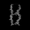
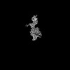
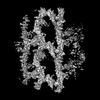
- PDB-9nu2: Uromodulin filament lattice in the straight arrangement from huma... -
+ Open data

- Basic information
| Entry | Database: PDB / ID: 9nu2 | ||||||
|---|---|---|---|---|---|---|---|
| Title | Uromodulin filament lattice in the straight arrangement from human urine | ||||||
 Components Components | Uromodulin | ||||||
 Keywords Keywords | ANTIMICROBIAL PROTEIN / Kidney / urine / uropathogen / filament / lattice / kidney disease / UTI / urinary tract infection / infection | ||||||
| Function / homology |  Function and homology information Function and homology informationcitric acid secretion / protein localization to extracellular region / metanephric thick ascending limb development / metanephric distal convoluted tubule development / connective tissue replacement / protein transport into plasma membrane raft / Asparagine N-linked glycosylation / organ or tissue specific immune response / collecting duct development / urea transmembrane transport ...citric acid secretion / protein localization to extracellular region / metanephric thick ascending limb development / metanephric distal convoluted tubule development / connective tissue replacement / protein transport into plasma membrane raft / Asparagine N-linked glycosylation / organ or tissue specific immune response / collecting duct development / urea transmembrane transport / micturition / metanephric ascending thin limb development / protein localization to vacuole / regulation of protein transport / juxtaglomerular apparatus development / intracellular chloride ion homeostasis / renal urate salt excretion / glomerular filtration / urate transport / antibacterial innate immune response / renal sodium ion absorption / neutrophil migration / response to water deprivation / regulation of urine volume / potassium ion homeostasis / intracellular sodium ion homeostasis / endoplasmic reticulum organization / intracellular phosphate ion homeostasis / heterophilic cell-cell adhesion / IgG binding / extrinsic component of membrane / leukocyte cell-cell adhesion / ciliary membrane / cellular response to unfolded protein / multicellular organismal response to stress / cellular defense response / renal water homeostasis / side of membrane / ERAD pathway / RNA splicing / tumor necrosis factor-mediated signaling pathway / lipid metabolic process / apoptotic signaling pathway / Golgi lumen / regulation of blood pressure / autophagy / intracellular calcium ion homeostasis / spindle pole / protein folding / response to lipopolysaccharide / gene expression / basolateral plasma membrane / defense response to Gram-negative bacterium / apical plasma membrane / cilium / response to xenobiotic stimulus / inflammatory response / negative regulation of cell population proliferation / calcium ion binding / cell surface / endoplasmic reticulum / : / extracellular exosome / membrane Similarity search - Function | ||||||
| Biological species |  Homo sapiens (human) Homo sapiens (human) | ||||||
| Method | ELECTRON MICROSCOPY / helical reconstruction / cryo EM / Resolution: 4.8 Å | ||||||
 Authors Authors | Chang, A.N. / Fitzpatrick, A.W.P. | ||||||
| Funding support | 1items
| ||||||
 Citation Citation | Journal: Structure / Year: 2025 Title: Structural basis of human uromodulin filament networks in uropathogen capture. Authors: Andrew N Chang / Gabriele Cerutti / Yuki Ogawa / Alia Basler / Willa Switzer / Mia Eng-Kohn / Carolyn Lee / Anthony W P Fitzpatrick /  Abstract: Uromodulin (UMOD), the most abundant protein in human urine, is essential for kidney function and urinary tract health. UMOD forms filaments that bind to uropathogenic bacteria, facilitating their ...Uromodulin (UMOD), the most abundant protein in human urine, is essential for kidney function and urinary tract health. UMOD forms filaments that bind to uropathogenic bacteria, facilitating their aggregation and clearance from the urinary tract. Here, we present the cryo-electron microscopy (cryo-EM) structure of the bacteria-binding D10C domain of UMOD and reveal its binding to the filament core. The details of D10C-core binding explain the formation of distinct filament lattice architectures adopted by UMOD. The D10C-core binding interface gives rise to diverse filament lattice structures, ranging from open and expansive to compact and dense conformations, or a combination of both. We hypothesize that other molecules present in urine may act as cross-linking agents, further stabilizing this binding interface and facilitating the connection of individual filaments into larger networks capable of effectively trapping bacteria. Structural mapping of kidney disease-related mutations points toward the abolition of disulfide bonds and promotion of mutant UMOD aggregation. | ||||||
| History |
|
- Structure visualization
| Structure viewer | Molecule: MolmilJmol/JSmol |
|---|
- Downloads & links
-Download
| PDBx/mmCIF format | 9nu2.cif.gz | 1 MB | Display | PDBx/mmCIF format |
|---|---|---|---|---|
| PDB format | pdb9nu2.ent.gz | Display | PDB format | |
| PDBx/mmJSON format | 9nu2.json.gz | Tree view | PDBx/mmJSON format | |
| Others |  Other downloads Other downloads |
-Validation report
| Arichive directory | https://data.pdbj.org/pub/pdb/validation_reports/nu/9nu2ftp://data.pdbj.org/pub/pdb/validation_reports/nu/9nu2 | HTTPS FTP |
|---|
-Related structure data
| Related structure data | 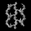 49793MC 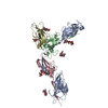 9nu1C 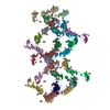 9nu3C M: map data used to model this data C: citing same article ( |
|---|---|
| Similar structure data |
-Links
 PDBj
PDBj


- Assembly
| Deposited unit | 
|
|---|---|
| 1 |
|
-Components
| #1: Protein | Mass: 69821.680 Da / Num. of mol.: 18 / Source method: isolated from a natural source / Source: (natural) Homo sapiens (human) / References: UniProt: P07911#2: Polysaccharide | 2-acetamido-2-deoxy-beta-D-glucopyranose-(1-4)-2-acetamido-2-deoxy-beta-D-glucopyranose Source method: isolated from a genetically manipulated source #3: Polysaccharide | alpha-D-mannopyranose-(1-3)-beta-D-mannopyranose-(1-4)-2-acetamido-2-deoxy-beta-D-glucopyranose-(1- ...alpha-D-mannopyranose-(1-3)-beta-D-mannopyranose-(1-4)-2-acetamido-2-deoxy-beta-D-glucopyranose-(1-4)-2-acetamido-2-deoxy-beta-D-glucopyranose Source method: isolated from a genetically manipulated source #4: Sugar | ChemComp-NAG /   Type: D-saccharide, beta linking / Mass: 221.208 Da / Num. of mol.: 15 / Source method: obtained synthetically / Formula: C8H15NO6 Type: D-saccharide, beta linking / Mass: 221.208 Da / Num. of mol.: 15 / Source method: obtained synthetically / Formula: C8H15NO6Has ligand of interest | N | Has protein modification | Y | |
|---|
-Experimental details
-Experiment
| Experiment | Method: ELECTRON MICROSCOPY |
|---|---|
| EM experiment | Aggregation state: FILAMENT / 3D reconstruction method: helical reconstruction |
- Sample preparation
| Component | Name: Uromodulin filament lattice in the straight arrangement from human urine Type: TISSUE / Entity ID: #1 / Source: NATURAL |
|---|---|
| Source (natural) | Organism: Homo sapiens (human) |
| Buffer solution | pH: 7.4 |
| Specimen | Embedding applied: NO / Shadowing applied: NO / Staining applied: NO / Vitrification applied: YES |
| Vitrification | Cryogen name: ETHANE |
- Electron microscopy imaging
| Experimental equipment |  Model: Titan Krios / Image courtesy: FEI Company |
|---|---|
| Microscopy | Model: TFS KRIOS |
| Electron gun | Electron source:  FIELD EMISSION GUN / Accelerating voltage: 300 kV / Illumination mode: FLOOD BEAM FIELD EMISSION GUN / Accelerating voltage: 300 kV / Illumination mode: FLOOD BEAM |
| Electron lens | Mode: BRIGHT FIELD / Nominal defocus max: 2500 nm / Nominal defocus min: 1200 nm |
| Image recording | Electron dose: 60 e/Å2 / Film or detector model: GATAN K3 BIOQUANTUM (6k x 4k) |
- Processing
| EM software | Name: PHENIX / Version: 1.21.2_5419 / Category: model refinement | ||||||||||||||||||||||||
|---|---|---|---|---|---|---|---|---|---|---|---|---|---|---|---|---|---|---|---|---|---|---|---|---|---|
| CTF correction | Type: NONE | ||||||||||||||||||||||||
| Helical symmerty | Angular rotation/subunit: 178.8 ° / Axial rise/subunit: 63.78 Å / Axial symmetry: C1 | ||||||||||||||||||||||||
| 3D reconstruction | Resolution: 4.8 Å / Resolution method: FSC 0.143 CUT-OFF / Num. of particles: 693160 / Symmetry type: HELICAL | ||||||||||||||||||||||||
| Refinement | Highest resolution: 4.8 Å Stereochemistry target values: REAL-SPACE (WEIGHTED MAP SUM AT ATOM CENTERS) | ||||||||||||||||||||||||
| Refine LS restraints |
|
