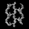
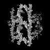
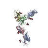
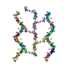
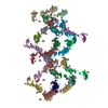
Journal: Structure / Year: 2025 Title: Structural basis of human uromodulin filament networks in uropathogen capture. Authors: Andrew N Chang / Gabriele Cerutti / Yuki Ogawa / Alia Basler / Willa Switzer / Mia Eng-Kohn / Carolyn Lee / Anthony W P Fitzpatrick / Abstract: Uromodulin (UMOD), the most abundant protein in human urine, is essential for kidney function and urinary tract health. UMOD forms filaments that bind to uropathogenic bacteria, facilitating their ...Uromodulin (UMOD), the most abundant protein in human urine, is essential for kidney function and urinary tract health. UMOD forms filaments that bind to uropathogenic bacteria, facilitating their aggregation and clearance from the urinary tract. Here, we present the cryo-electron microscopy (cryo-EM) structure of the bacteria-binding D10C domain of UMOD and reveal its binding to the filament core. The details of D10C-core binding explain the formation of distinct filament lattice architectures adopted by UMOD. The D10C-core binding interface gives rise to diverse filament lattice structures, ranging from open and expansive to compact and dense conformations, or a combination of both. We hypothesize that other molecules present in urine may act as cross-linking agents, further stabilizing this binding interface and facilitating the connection of individual filaments into larger networks capable of effectively trapping bacteria. Structural mapping of kidney disease-related mutations points toward the abolition of disulfide bonds and promotion of mutant UMOD aggregation.
In the structure databanks used in Yorodumi, some data are registered as the other names, "COVID-19 virus" and "2019-nCoV". Here are the details of the virus and the list of structure data.
Jan 31, 2019. EMDB accession codes are about to change! (news from PDBe EMDB page)
EMDB accession codes are about to change! (news from PDBe EMDB page)
The allocation of 4 digits for EMDB accession codes will soon come to an end. Whilst these codes will remain in use, new EMDB accession codes will include an additional digit and will expand incrementally as the available range of codes is exhausted. The current 4-digit format prefixed with “EMD-” (i.e. EMD-XXXX) will advance to a 5-digit format (i.e. EMD-XXXXX), and so on. It is currently estimated that the 4-digit codes will be depleted around Spring 2019, at which point the 5-digit format will come into force.
The EM Navigator/Yorodumi systems omit the EMD- prefix.
Related info.:Q: What is EMD? / ID/Accession-code notation in Yorodumi/EM Navigator
Yorodumi is a browser for structure data from EMDB, PDB, SASBDB, etc.
This page is also the successor to EM Navigator detail page, and also detail information page/front-end page for Omokage search.
The word "yorodu" (or yorozu) is an old Japanese word meaning "ten thousand". "mi" (miru) is to see.
Related info.:EMDB / PDB / SASBDB / Comparison of 3 databanks / Yorodumi Search / Aug 31, 2016. New EM Navigator & Yorodumi / Yorodumi Papers / Jmol/JSmol / Function and homology information / Changes in new EM Navigator and Yorodumi
 Movie
Movie Controller

 Yorodumi
 Open data
 Basic information
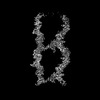
 Map data
 Sample
 Keywords
 Homo sapiens (human)
 Authors
 Citation
 Structure visualization
 Downloads & links
 EMDB map data format
 emd_49752.png
 http://ftp.pdbj.org/pub/emdb/structures/EMD-49752
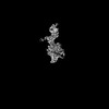
 Z (Sec.)
 Y (Row.)
 X (Col.)


 Sample components
 Processing
 Electron microscopy
 FIELD EMISSION GUN
