 Movie
Movie Controller


+ Open data

- Basic information
| Entry | Database: PDB / ID: 9ntm | |||||||||
|---|---|---|---|---|---|---|---|---|---|---|
| Title | SPEF1 bound to 14-pf microtubule | |||||||||
 Components Components |
| |||||||||
 Keywords Keywords | PROTEIN BINDING / microtubules / spef1 / seam | |||||||||
| Function / homology |  Function and homology information Function and homology informationaxonemal central apparatus / axonemal central apparatus assembly / regulation of Wnt signaling pathway, planar cell polarity pathway / 9+2 motile cilium / cilium movement / filopodium assembly / negative regulation of microtubule depolymerization / microtubule bundle formation / lamellipodium assembly / regulation of cytoskeleton organization ...axonemal central apparatus / axonemal central apparatus assembly / regulation of Wnt signaling pathway, planar cell polarity pathway / 9+2 motile cilium / cilium movement / filopodium assembly / negative regulation of microtubule depolymerization / microtubule bundle formation / lamellipodium assembly / regulation of cytoskeleton organization / microvillus / host cell membrane / axoneme / cellular response to interleukin-4 / microtubule-based process / cytoplasmic microtubule / ciliary tip / stress fiber / bioluminescence / filopodium / generation of precursor metabolites and energy / microtubule cytoskeleton organization / structural constituent of cytoskeleton / microtubule cytoskeleton / mitotic cell cycle / cell migration / lamellipodium / double-stranded RNA binding / actin binding / cilium / microtubule binding / basolateral plasma membrane / microtubule / Hydrolases; Acting on acid anhydrides; Acting on GTP to facilitate cellular and subcellular movement / apical plasma membrane / GTPase activity / viral envelope / ubiquitin protein ligase binding / symbiont entry into host cell / virion attachment to host cell / GTP binding / virion membrane / metal ion binding / cytosol / cytoplasm Similarity search - Function | |||||||||
| Biological species |  Homo sapiens (human) Homo sapiens (human) Recombinant vesicular stomatitis Indiana virus rVSV-G/GFP Recombinant vesicular stomatitis Indiana virus rVSV-G/GFP | |||||||||
| Method | ELECTRON MICROSCOPY / subtomogram averaging / cryo EM / Resolution: 7.1 Å | |||||||||
 Authors Authors | Legal, T. / Bui, K.H. | |||||||||
| Funding support |  Canada, 2items Canada, 2items
| |||||||||
 Citation Citation | Journal: Curr Biol / Year: 2025 Title: Structure of the ciliary tip central pair reveals the unique role of the microtubule-seam binding protein SPEF1. Authors: Thibault Legal / Ewa Joachimiak / Mireya Parra / Wang Peng / Amanda Tam / Corbin Black / Mayukh Guha / Chau Anh Nguyen / Avrin Ghanaeian / Melissa Valente-Paterno / Gary Brouhard / Jacek ...Authors: Thibault Legal / Ewa Joachimiak / Mireya Parra / Wang Peng / Amanda Tam / Corbin Black / Mayukh Guha / Chau Anh Nguyen / Avrin Ghanaeian / Melissa Valente-Paterno / Gary Brouhard / Jacek Gaertig / Dorota Wloga / Khanh Huy Bui /   Abstract: Motile cilia are unique organelles with the ability to move autonomously. The force generated by beating cilia propels cells and moves fluids. The ciliary skeleton is made of peripheral doublet ...Motile cilia are unique organelles with the ability to move autonomously. The force generated by beating cilia propels cells and moves fluids. The ciliary skeleton is made of peripheral doublet microtubules and a central pair (CP) with a distinct structure at the tip. In this study, we present a high-resolution structure of the CP in the ciliary tip of the ciliate Tetrahymena thermophila and identify several tip proteins that bind and form unique patterns on both microtubules of the tip CP. Two of those proteins that contain tubulin polymerization-promoting protein (TPPP)-like domains, TLP1 and TLP2, bind to high curvature regions of the microtubule. TLP2, which contains two TPPP-like domains, is an unusually long protein that wraps laterally around half a microtubule and forms the bridge between the two microtubules. Moreover, we found that the conserved protein SPEF1 binds to both microtubule seams and crosslinked the two microtubules. In vitro, human SPEF1 binds to the microtubule seam as visualized by cryoelectron tomography and subtomogram averaging. Single-molecule microtubule dynamics assays indicate that SPEF1 stabilizes microtubules in vitro. Together, these data show that the proteins in the tip CP maintain stable microtubule structures and play important roles in maintaining the integrity of the axoneme. | |||||||||
| History |
|
- Structure visualization
| Structure viewer | Molecule: MolmilJmol/JSmol |
|---|
- Downloads & links
-Download
| PDBx/mmCIF format | 9ntm.cif.gz | 6.1 MB | Display | PDBx/mmCIF format |
|---|---|---|---|---|
| PDB format | pdb9ntm.ent.gz | Display | PDB format | |
| PDBx/mmJSON format | 9ntm.json.gz | Tree view | PDBx/mmJSON format | |
| Others |  Other downloads Other downloads |
-Validation report
| Arichive directory | https://data.pdbj.org/pub/pdb/validation_reports/nt/9ntmftp://data.pdbj.org/pub/pdb/validation_reports/nt/9ntm | HTTPS FTP |
|---|
-Related structure data
| Related structure data | 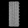 49760MC  9nw3C  9ot2C M: map data used to model this data C: citing same article ( |
|---|---|
| Similar structure data |


-Links
 PDBj
PDBj


- Assembly
| Deposited unit | 
|
|---|---|
| 1 |
|
-Components
-Protein , 3 types, 89 molecules 1A1B1C1D1EAAACAEBABCBECACCCEDADCDEEAECEEFAFCFEGAGCGEHAHCHEIA...
| #1: Protein | Mass: 44324.262 Da / Num. of mol.: 5 Source method: isolated from a genetically manipulated source Source: (gene. exp.) Homo sapiens (human), (gene. exp.) Recombinant vesicular stomatitis Indiana virus rVSV-G/GFPGene: SPEF1, C20orf28, G, GFP / Production host:  #2: Protein | Mass: 50204.445 Da / Num. of mol.: 42 / Source method: isolated from a natural source / Source: (natural) #3: Protein | Mass: 49983.824 Da / Num. of mol.: 42 / Source method: isolated from a natural source / Source: (natural) |
|---|
-Non-polymers , 4 types, 168 molecules 


| #4: Chemical | ChemComp-GTP /  Mass: 523.180 Da / Num. of mol.: 42 / Source method: obtained synthetically / Formula: C10H16N5O14P3 / Comment: GTP, energy-carrying molecule*YM Mass: 523.180 Da / Num. of mol.: 42 / Source method: obtained synthetically / Formula: C10H16N5O14P3 / Comment: GTP, energy-carrying molecule*YM#5: Chemical | ChemComp-MG /  Mass: 24.305 Da / Num. of mol.: 42 / Source method: obtained synthetically / Formula: Mg Mass: 24.305 Da / Num. of mol.: 42 / Source method: obtained synthetically / Formula: Mg#6: Chemical | ChemComp-GDP /  Type: RNA linking / Mass: 443.201 Da / Num. of mol.: 42 / Source method: obtained synthetically / Formula: C10H15N5O11P2 / Comment: GDP, energy-carrying molecule*YM Type: RNA linking / Mass: 443.201 Da / Num. of mol.: 42 / Source method: obtained synthetically / Formula: C10H15N5O11P2 / Comment: GDP, energy-carrying molecule*YM#7: Chemical | ChemComp-TA1 /  Mass: 853.906 Da / Num. of mol.: 42 / Source method: obtained synthetically / Formula: C47H51NO14 / Comment: medication, chemotherapy*YM Mass: 853.906 Da / Num. of mol.: 42 / Source method: obtained synthetically / Formula: C47H51NO14 / Comment: medication, chemotherapy*YM |
|---|
-Details
| Has ligand of interest | N |
|---|---|
| Has protein modification | N |
-Experimental details
-Experiment
| Experiment | Method: ELECTRON MICROSCOPY |
|---|---|
| EM experiment | Aggregation state: FILAMENT / 3D reconstruction method: subtomogram averaging |
- Sample preparation
| Component | Name: 14-protofilament microtubule bound by SPEF1 CH-domain / Type: COMPLEX / Entity ID: #2-#3, #1 / Source: MULTIPLE SOURCES | ||||||||||||
|---|---|---|---|---|---|---|---|---|---|---|---|---|---|
| Molecular weight | Experimental value: NO | ||||||||||||
| Source (natural) |
| ||||||||||||
| Source (recombinant) | Organism: | ||||||||||||
| Buffer solution | pH: 6.8 | ||||||||||||
| Specimen | Embedding applied: NO / Shadowing applied: NO / Staining applied: NO / Vitrification applied: YES | ||||||||||||
| Vitrification | Cryogen name: ETHANE |
- Electron microscopy imaging
| Experimental equipment |  Model: Titan Krios / Image courtesy: FEI Company |
|---|---|
| Microscopy | Model: TFS KRIOS |
| Electron gun | Electron source:  FIELD EMISSION GUN / Accelerating voltage: 300 kV / Illumination mode: FLOOD BEAM FIELD EMISSION GUN / Accelerating voltage: 300 kV / Illumination mode: FLOOD BEAM |
| Electron lens | Mode: BRIGHT FIELD / Nominal defocus max: 4000 nm / Nominal defocus min: 2000 nm |
| Image recording | Electron dose: 4 e/Å2 / Avg electron dose per subtomogram: 164 e/Å2 / Film or detector model: GATAN K3 (6k x 4k) |
- Processing
| EM software | Name: PHENIX / Version: 1.21_5207 / Category: model refinement |
|---|---|
| CTF correction | Type: PHASE FLIPPING AND AMPLITUDE CORRECTION |
| 3D reconstruction | Resolution: 7.1 Å / Resolution method: FSC 0.143 CUT-OFF / Num. of particles: 9625 / Symmetry type: POINT |
| EM volume selection | Num. of tomograms: 58 / Num. of volumes extracted: 20342 |
