Natural Sciences and Engineering Research Council (NSERC, Canada)
RGPIN-2022-04774
Canada
Citation
Journal: Curr Biol / Year: 2025 Title: Structure of the ciliary tip central pair reveals the unique role of the microtubule-seam binding protein SPEF1. Authors: Thibault Legal / Ewa Joachimiak / Mireya Parra / Wang Peng / Amanda Tam / Corbin Black / Mayukh Guha / Chau Anh Nguyen / Avrin Ghanaeian / Melissa Valente-Paterno / Gary Brouhard / Jacek ...Authors: Thibault Legal / Ewa Joachimiak / Mireya Parra / Wang Peng / Amanda Tam / Corbin Black / Mayukh Guha / Chau Anh Nguyen / Avrin Ghanaeian / Melissa Valente-Paterno / Gary Brouhard / Jacek Gaertig / Dorota Wloga / Khanh Huy Bui / Abstract: Motile cilia are unique organelles with the ability to move autonomously. The force generated by beating cilia propels cells and moves fluids. The ciliary skeleton is made of peripheral doublet ...Motile cilia are unique organelles with the ability to move autonomously. The force generated by beating cilia propels cells and moves fluids. The ciliary skeleton is made of peripheral doublet microtubules and a central pair (CP) with a distinct structure at the tip. In this study, we present a high-resolution structure of the CP in the ciliary tip of the ciliate Tetrahymena thermophila and identify several tip proteins that bind and form unique patterns on both microtubules of the tip CP. Two of those proteins that contain tubulin polymerization-promoting protein (TPPP)-like domains, TLP1 and TLP2, bind to high curvature regions of the microtubule. TLP2, which contains two TPPP-like domains, is an unusually long protein that wraps laterally around half a microtubule and forms the bridge between the two microtubules. Moreover, we found that the conserved protein SPEF1 binds to both microtubule seams and crosslinked the two microtubules. In vitro, human SPEF1 binds to the microtubule seam as visualized by cryoelectron tomography and subtomogram averaging. Single-molecule microtubule dynamics assays indicate that SPEF1 stabilizes microtubules in vitro. Together, these data show that the proteins in the tip CP maintain stable microtubule structures and play important roles in maintaining the integrity of the axoneme.
In the structure databanks used in Yorodumi, some data are registered as the other names, "COVID-19 virus" and "2019-nCoV". Here are the details of the virus and the list of structure data.
Jan 31, 2019. EMDB accession codes are about to change! (news from PDBe EMDB page)
EMDB accession codes are about to change! (news from PDBe EMDB page)
The allocation of 4 digits for EMDB accession codes will soon come to an end. Whilst these codes will remain in use, new EMDB accession codes will include an additional digit and will expand incrementally as the available range of codes is exhausted. The current 4-digit format prefixed with “EMD-” (i.e. EMD-XXXX) will advance to a 5-digit format (i.e. EMD-XXXXX), and so on. It is currently estimated that the 4-digit codes will be depleted around Spring 2019, at which point the 5-digit format will come into force.
The EM Navigator/Yorodumi systems omit the EMD- prefix.
Related info.:Q: What is EMD? / ID/Accession-code notation in Yorodumi/EM Navigator
Yorodumi is a browser for structure data from EMDB, PDB, SASBDB, etc.
This page is also the successor to EM Navigator detail page, and also detail information page/front-end page for Omokage search.
The word "yorodu" (or yorozu) is an old Japanese word meaning "ten thousand". "mi" (miru) is to see.
Related info.:EMDB / PDB / SASBDB / Comparison of 3 databanks / Yorodumi Search / Aug 31, 2016. New EM Navigator & Yorodumi / Yorodumi Papers / Jmol/JSmol / Function and homology information / Changes in new EM Navigator and Yorodumi
 Movie
Movie Controller


 Open data
 Basic information
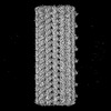
 Map data
 Sample
 Keywords
 Function and homology information
 Homo sapiens (human) /
  Recombinant vesicular stomatitis Indiana virus rVSV-G/GFP /
 Authors
 Canada, 2 items
  Citation

 Structure visualization
 Downloads & links
 emd_49760.png
 http://ftp.pdbj.org/pub/emdb/structures/EMD-49760


 Z (Sec.)
 Y (Row.)
 X (Col.)


 Sample components


 Processing
 Electron microscopy
 FIELD EMISSION GUN
