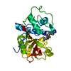
ジャーナル: bioRxiv / 年: 2025 タイトル: Combining MicroED and native mass spectrometry for structural discovery of enzyme-biosynthetic inhibitor complexes. 著者: Niko W Vlahakis / Cameron W Flowers / Mengting Liu / Matthew Agdanowski / Samuel Johnson / Jacob A Summers / Catherine Keyser / Phoebe Russell / Samuel Rose / Julien Orlans / Nima Adhami / Yu ...著者: Niko W Vlahakis / Cameron W Flowers / Mengting Liu / Matthew Agdanowski / Samuel Johnson / Jacob A Summers / Catherine Keyser / Phoebe Russell / Samuel Rose / Julien Orlans / Nima Adhami / Yu Chen / Michael R Sawaya / Shibom Basu / Daniele de Sanctis / Soichi Wakatsuki / Hosea M Nelson / Joseph A Loo / Yi Tang / Jose A Rodriguez / 要旨: With the goal of accelerating the discovery of small molecule-protein complexes, we leverage fast, low-dose, event based electron counting microcrystal electron diffraction (MicroED) data collection ...With the goal of accelerating the discovery of small molecule-protein complexes, we leverage fast, low-dose, event based electron counting microcrystal electron diffraction (MicroED) data collection and native mass spectrometry. This approach resolves structures of the epoxide-based cysteine protease inhibitor, and natural product, E-64, and its biosynthetic analogs bound to the model cysteine protease, papain. The combined structural power of MicroED and the analytical capabilities of native mass spectrometry (ED-MS) allows assignment of papain structures bound to E-64-like ligands with data obtained from crystal slurries soaked with mixtures of known inhibitors, and crude biosynthetic reactions. ED-MS further discriminates the highest-affinity ligand soaked into microcrystals from a broad inhibitor cocktail, and identifies multiple similarly high-affinity ligands soaked into microcrystals simultaneously. This extends to libraries of printed ligands dispensed directly onto TEM grids and later soaked with papain microcrystal slurries. ED-MS identifies papain binding to its preferred natural products, by showing that two analogues of E-64 outcompete others in binding to papain crystals, and by detecting papain bound to E-64 and an analogue from crude biosynthetic reactions, without purification. This illustrates the utility of ED-MS for natural product ligand discovery and for structure-based screening of small molecule binders to macromolecular targets.
 ムービー
ムービー コントローラー


 データを開く
 基本情報
基本情報 要素
要素 キーワード
キーワード 機能・相同性情報
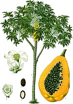 Carica papaya (パパイア)
 X線回折 /
  データ登録者
 米国, 3件
  引用
引用
 構造の表示
構造の表示 ダウンロードとリンク
 その他のダウンロード


 PDBj
PDBj
 集合体
集合体
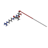
 分子量: 360.429 Da / 分子数: 1 / 由来タイプ: 合成 / 式: C15H30N5O5 / タイプ: SUBJECT OF INVESTIGATION
 分子量: 18.015 Da / 分子数: 223 / 由来タイプ: 天然 / 式: H2O
 試料調製
試料調製 解析
解析