 Movie
Movie Controller


+ Open data

- Basic information
| Entry | Database: PDB / ID: 9na8 | ||||||
|---|---|---|---|---|---|---|---|
| Title | Augmin1345 Extended-body | ||||||
 Components Components |
| ||||||
 Keywords Keywords | PLANT PROTEIN | ||||||
| Function / homology |  Function and homology information Function and homology informationphragmoplast microtubule organization / HAUS complex / microtubule minus-end binding / phragmoplast / microtubule organizing center organization / microtubule organizing center / spindle assembly / bioluminescence / spindle microtubule / generation of precursor metabolites and energy ...phragmoplast microtubule organization / HAUS complex / microtubule minus-end binding / phragmoplast / microtubule organizing center organization / microtubule organizing center / spindle assembly / bioluminescence / spindle microtubule / generation of precursor metabolites and energy / spindle / mitotic spindle / microtubule / cell division / nucleus / cytosol Similarity search - Function | ||||||
| Biological species |    Aequorea victoria (jellyfish) Aequorea victoria (jellyfish) | ||||||
| Method | ELECTRON MICROSCOPY / single particle reconstruction / cryo EM / Resolution: 3.5 Å | ||||||
 Authors Authors | Ashaduzzaman, M. / Al-Bassam, J. / Taheri, A. | ||||||
| Funding support |  United States, 1items United States, 1items
| ||||||
 Citation Citation | Journal: bioRxiv / Year: 2025 Title: Cryo-EM structures of the Plant Augmin reveal its intertwined coiled-coil assembly, antiparallel dimerization and NEDD1 binding mechanisms. Authors: Md Ashaduzzaman / Aryan Taheri / Yuh-Ru Julie Lee / Yuqi Tang / Fei Guo / Stephen D Fried / Bo Liu / Jawdat Al-Bassam / Abstract: Microtubule (MT) branch nucleation is fundamental for building parallel MT networks in eukaryotic cells. In plants and metazoans, MT branch nucleation requires Augmin and NEDD1 proteins which bind ...Microtubule (MT) branch nucleation is fundamental for building parallel MT networks in eukaryotic cells. In plants and metazoans, MT branch nucleation requires Augmin and NEDD1 proteins which bind along MTs and then recruit and activate the gamma-tubulin ring complex (γ-TuRC). Augmin is a fork-shaped assembly composed of eight coiled-coil subunits, while NEDD1 is a WD40 β-propellor protein that bridges across MTs, Augmin, and γ-TuRC during MT branch nucleation. Here, we reconstitute hetero-tetrameric and hetero-octameric Arabidopsis thaliana Augmin assemblies, resolve their subunit interactions using crosslinking mass spectrometry and determine 3.7 to 7.3-Å cryo-EM structures for the V-junction and extended regions of Augmin. These structures allowed us to generate a complete de novo plant Augmin model that reveals the long-range multi coiled-coil interfaces that stabilize its 40-nm hetero-octameric fork-shaped organization. We discovered the dual calponin homology (CH) domain forming its MT binding site at the end of its V-junction undertake open and closed conformations. We determined a 12-Å dimeric Augmin cryo-EM structure revealing Augmin undergoes anti-parallel dimerization through two conserved surfaces along Augmin's extended region. We reconstituted the NEDD1 WD40 β-propellor with Augmin revealing it directly binds on top its V-junction and enhances Augmin dimerization. Our studies suggest that cooperativity between the Augmin dual CH domains and NEDD1 WD40 binding site may regulate Augmin V-junction dual binding to MT lattices. This unique V-shaped dual binding and organization anchors Augmins along MTs generating a platform to recruit γ-TuRC and activate branched MT nucleation. | ||||||
| History |
|
- Structure visualization
| Structure viewer | Molecule: MolmilJmol/JSmol |
|---|
- Downloads & links
-Download
| PDBx/mmCIF format | 9na8.cif.gz | 406.8 KB | Display | PDBx/mmCIF format |
|---|---|---|---|---|
| PDB format | pdb9na8.ent.gz | 264.4 KB | Display | PDB format |
| PDBx/mmJSON format | 9na8.json.gz | Tree view | PDBx/mmJSON format | |
| Others |  Other downloads Other downloads |
-Validation report
| Arichive directory | https://data.pdbj.org/pub/pdb/validation_reports/na/9na8ftp://data.pdbj.org/pub/pdb/validation_reports/na/9na8 | HTTPS FTP |
|---|
-Related structure data
| Related structure data |  49182MC 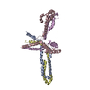 9na9C  9nbaC 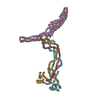 9nbbC 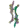 9nbdC M: map data used to model this data C: citing same article ( |
|---|---|
| Similar structure data |


-Links
 PDBj
PDBj

- Assembly
| Deposited unit | 
|
|---|---|
| 1 |
|
-Components
| #1: Protein | Mass: 33537.996 Da / Num. of mol.: 1 Source method: isolated from a genetically manipulated source Source: (gene. exp.)  |
|---|---|
| #2: Protein | Mass: 47825.855 Da / Num. of mol.: 1 Source method: isolated from a genetically manipulated source Source: (gene. exp.) |
| #3: Protein | Mass: 116629.992 Da / Num. of mol.: 1 Source method: isolated from a genetically manipulated source Source: (gene. exp.) Aequorea victoria (jellyfish)Gene: AUG5, At5g38880, K15E6.9, GFP / Production host: |
| #4: Protein | Mass: 69809.453 Da / Num. of mol.: 1 Source method: isolated from a genetically manipulated source Source: (gene. exp.) |
| Has protein modification | Y |
-Experimental details
-Experiment
| Experiment | Method: ELECTRON MICROSCOPY |
|---|---|
| EM experiment | Aggregation state: PARTICLE / 3D reconstruction method: single particle reconstruction |
- Sample preparation
| Component | Name: Augmin1,3,4,5 Tetramer extended complex / Type: COMPLEX Details: AUG1,3,4,5 construct was assembled into a polycistronic expression vector with an AUG5 C-terminal GFP and His Entity ID: all / Source: RECOMBINANT |
|---|---|
| Molecular weight | Value: 0.31 MDa / Experimental value: YES |
| Source (natural) | Organism: |
| Source (recombinant) | Organism: |
| Buffer solution | pH: 7.5 Details: 50mM HEPES,150mM KCl,1mM EGTA,1mM MgCl2,10mM betamercaptoethanol |
| Buffer component | Conc.: 50 mM / Name: HEPES |
| Specimen | Conc.: 1 mg/ml / Embedding applied: NO / Shadowing applied: NO / Staining applied: NO / Vitrification applied: YES / Details: Gel filtration purified |
| Specimen support | Grid material: COPPER / Grid mesh size: 300 divisions/in. / Grid type: Quantifoil R1.2/1.3 |
| Vitrification | Instrument: LEICA EM GP / Cryogen name: ETHANE / Humidity: 95 % / Chamber temperature: 293 K |
- Electron microscopy imaging
| Microscopy | Model: TFS GLACIOS |
|---|---|
| Electron gun | Electron source:  FIELD EMISSION GUN / Accelerating voltage: 200 kV / Illumination mode: FLOOD BEAM FIELD EMISSION GUN / Accelerating voltage: 200 kV / Illumination mode: FLOOD BEAM |
| Electron lens | Mode: BRIGHT FIELD / Nominal defocus max: 1800 nm / Nominal defocus min: 600 nm |
| Image recording | Electron dose: 60 e/Å2 / Film or detector model: GATAN K3 (6k x 4k) |
- Processing
| EM software |
| ||||||||||||||||||||||||||||
|---|---|---|---|---|---|---|---|---|---|---|---|---|---|---|---|---|---|---|---|---|---|---|---|---|---|---|---|---|---|
| CTF correction | Type: PHASE FLIPPING AND AMPLITUDE CORRECTION | ||||||||||||||||||||||||||||
| 3D reconstruction | Resolution: 3.5 Å / Resolution method: FSC 0.143 CUT-OFF / Num. of particles: 72002 / Num. of class averages: 1 / Symmetry type: POINT | ||||||||||||||||||||||||||||
| Refinement | Cross valid method: NONE Stereochemistry target values: GeoStd + Monomer Library + CDL v1.2 | ||||||||||||||||||||||||||||
| Displacement parameters | Biso mean: 136.84 Å2 | ||||||||||||||||||||||||||||
| Refine LS restraints |
|
