 Movie
Movie Controller

[English] 日本語
 Yorodumi
- PDB-9mfm: Structure of zebrafish OTOP1 in nanodisc in complex with inhibito... -
+ Open data

- Basic information
| Entry | Database: PDB / ID: 9mfm | ||||||
|---|---|---|---|---|---|---|---|
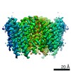
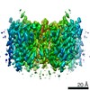
| Title | Structure of zebrafish OTOP1 in nanodisc in complex with inhibitor C2.36 | ||||||
 Components Components | Proton channel OTOP1 | ||||||
 Keywords Keywords | MEMBRANE PROTEIN / proton channel / ion channel / OTOP / Otopetrin | ||||||
| Function / homology |  Function and homology information Function and homology informationotolith formation / otolith mineralization / proton channel activity / inner ear morphogenesis / cholesterol binding / negative regulation of type II interferon-mediated signaling pathway / microvillus / proton transmembrane transport / cellular response to insulin stimulus / membrane ...otolith formation / otolith mineralization / proton channel activity / inner ear morphogenesis / cholesterol binding / negative regulation of type II interferon-mediated signaling pathway / microvillus / proton transmembrane transport / cellular response to insulin stimulus / membrane / identical protein binding / plasma membrane Similarity search - Function | ||||||
| Biological species |  | ||||||
| Method | ELECTRON MICROSCOPY / single particle reconstruction / cryo EM / Resolution: 3.42 Å | ||||||
 Authors Authors | Burendei, B. / Ward, A.B. | ||||||
| Funding support |  United States, 1items United States, 1items
| ||||||
 Citation Citation | Journal: Nat Commun / Year: 2025 Title: Structure-guided discovery of Otopetrin 1 inhibitors reveals druggable binding sites at the intrasubunit interface. Authors: Batuujin Burendei / Joshua P Kaplan / Gerardo M Orellana / Emily R Liman / Stefano Forli / Andrew B Ward / Abstract: Proton conductance across cell membranes serves many biological functions, ranging from the regulation of intracellular and extracellular pH to the generation of electrical signals that lead to ...Proton conductance across cell membranes serves many biological functions, ranging from the regulation of intracellular and extracellular pH to the generation of electrical signals that lead to sour taste perception. Otopetrins (OTOPs) are a conserved, eukaryotic family of proton-selective ion channels, one of which (OTOP1) serves as a gustatory sensor for sour tastes and ammonium chloride. As the functional properties and structures of OTOP channels were only recently described, there are presently few tools available to modulate their activity. Here, we perform subsequent rounds of molecular docking-based virtual screening against the structure of zebrafish OTOP1, followed by functional testing using whole-cell patch-clamp electrophysiology, and identify several small molecule inhibitors that are effective in the low-to-mid µM range. Cryo-electron microscopy structures reveal inhibitor binding sites in the intrasubunit interface that are validated by functional testing of mutant channels. Our findings reveal pockets that can be targeted for small molecule discovery to develop modulators for Otopetrins. Such modulators can serve as useful toolkit molecules for future investigations of structure-function relationships or physiological roles of Otopetrins. | ||||||
| History |
|
- Structure visualization
| Structure viewer | Molecule: MolmilJmol/JSmol |
|---|
- Downloads & links
-Download
| PDBx/mmCIF format | 9mfm.cif.gz | 169.7 KB | Display | PDBx/mmCIF format |
|---|---|---|---|---|
| PDB format | pdb9mfm.ent.gz | Display | PDB format | |
| PDBx/mmJSON format | 9mfm.json.gz | Tree view | PDBx/mmJSON format | |
| Others |  Other downloads Other downloads |
-Validation report
| Arichive directory | https://data.pdbj.org/pub/pdb/validation_reports/mf/9mfmftp://data.pdbj.org/pub/pdb/validation_reports/mf/9mfm | HTTPS FTP |
|---|
-Related structure data
| Related structure data |  48235MC  9mffC  9mflC C: citing same article ( M: map data used to model this data |
|---|---|
| Similar structure data |


-Links
 PDBj
PDBj


- Assembly
| Deposited unit | 
|
|---|---|
| 1 |
|
-Components
| #1: Protein | Mass: 65817.000 Da / Num. of mol.: 2 Source method: isolated from a genetically manipulated source Details: first two residues (GP) are leftover residues from a 3C protease cleavage site. 3rd residue (V) corresponds to the 2nd residue of the zebrafish OTOP1 sequence (Uniprot:Q7ZWK8) Source: (gene. exp.)  Homo sapiens (human) / References: UniProt: Q7ZWK8 Homo sapiens (human) / References: UniProt: Q7ZWK8#2: Chemical | ChemComp-Y01 /   Mass: 486.726 Da / Num. of mol.: 4 / Source method: obtained synthetically / Formula: C31H50O4 Mass: 486.726 Da / Num. of mol.: 4 / Source method: obtained synthetically / Formula: C31H50O4#3: Chemical |   Mass: 386.654 Da / Num. of mol.: 2 / Source method: obtained synthetically / Formula: C27H46O Mass: 386.654 Da / Num. of mol.: 2 / Source method: obtained synthetically / Formula: C27H46O#4: Chemical | ChemComp-A1BKT / Mass: 279.293 Da / Num. of mol.: 6 / Source method: obtained synthetically / Formula: C16H13N3O2 / Feature type: SUBJECT OF INVESTIGATION Has ligand of interest | Y | Has protein modification | N | |
|---|
-Experimental details
-Experiment
| Experiment | Method: ELECTRON MICROSCOPY |
|---|---|
| EM experiment | Aggregation state: PARTICLE / 3D reconstruction method: single particle reconstruction |
- Sample preparation
| Component | Name: zebrafish OTOP1 in MSP2N2 nanodisc / Type: COMPLEX / Entity ID: #1 / Source: RECOMBINANT | ||||||||||||||||||||
|---|---|---|---|---|---|---|---|---|---|---|---|---|---|---|---|---|---|---|---|---|---|
| Molecular weight | Value: 0.131 MDa / Experimental value: NO | ||||||||||||||||||||
| Source (natural) | Organism: | ||||||||||||||||||||
| Source (recombinant) | Organism: Homo sapiens (human) / Cell: HEK293F / Plasmid: pEG | ||||||||||||||||||||
| Buffer solution | pH: 8 | ||||||||||||||||||||
| Buffer component |
| ||||||||||||||||||||
| Specimen | Conc.: 5.3 mg/ml / Embedding applied: NO / Shadowing applied: NO / Staining applied: NO / Vitrification applied: YES Details: sample was complexed with small molecule C2.36, reaching a final concentration of 1 mM, and 5% DMSO | ||||||||||||||||||||
| Specimen support | Grid material: GOLD / Grid mesh size: 300 divisions/in. / Grid type: UltrAuFoil R1.2/1.3 | ||||||||||||||||||||
| Vitrification | Instrument: FEI VITROBOT MARK IV / Cryogen name: ETHANE / Humidity: 100 % / Chamber temperature: 283.15 K |
- Electron microscopy imaging
| Microscopy | Model: TFS GLACIOS |
|---|---|
| Electron gun | Electron source:  FIELD EMISSION GUN / Accelerating voltage: 200 kV / Illumination mode: FLOOD BEAM FIELD EMISSION GUN / Accelerating voltage: 200 kV / Illumination mode: FLOOD BEAM |
| Electron lens | Mode: BRIGHT FIELD / Nominal magnification: 190000 X / Nominal defocus max: 1800 nm / Nominal defocus min: 800 nm / Cs: 2.7 mm / C2 aperture diameter: 70 µm / Alignment procedure: COMA FREE |
| Specimen holder | Cryogen: NITROGEN / Specimen holder model: OTHER |
| Image recording | Average exposure time: 3.5 sec. / Electron dose: 45 e/Å2 / Film or detector model: TFS FALCON 4i (4k x 4k) / Num. of grids imaged: 3 / Num. of real images: 26378 |
| Image scans | Width: 4096 / Height: 4096 |
- Processing
| EM software | Name: PHENIX / Version: 1.20.1_4487: / Category: model refinement |
|---|---|
| Image processing | Details: Micrographs with CTF estimates (cryoSPARC Live) worse than 5A were discarded. |
| CTF correction | Type: PHASE FLIPPING AND AMPLITUDE CORRECTION |
| Particle selection | Num. of particles selected: 7236014 |
| Symmetry | Point symmetry: C2 (2 fold cyclic) |
| 3D reconstruction | Resolution: 3.42 Å / Resolution method: FSC 0.143 CUT-OFF / Num. of particles: 290491 Details: final reported resolution of 3.42A was obtained through the Remote 3DFSC Processing server Num. of class averages: 2 / Symmetry type: POINT |
| Atomic model building | Protocol: AB INITIO MODEL / Space: REAL |
| Atomic model building | PDB-ID: 6NF4 Accession code: 6NF4 / Source name: PDB / Type: experimental model |
